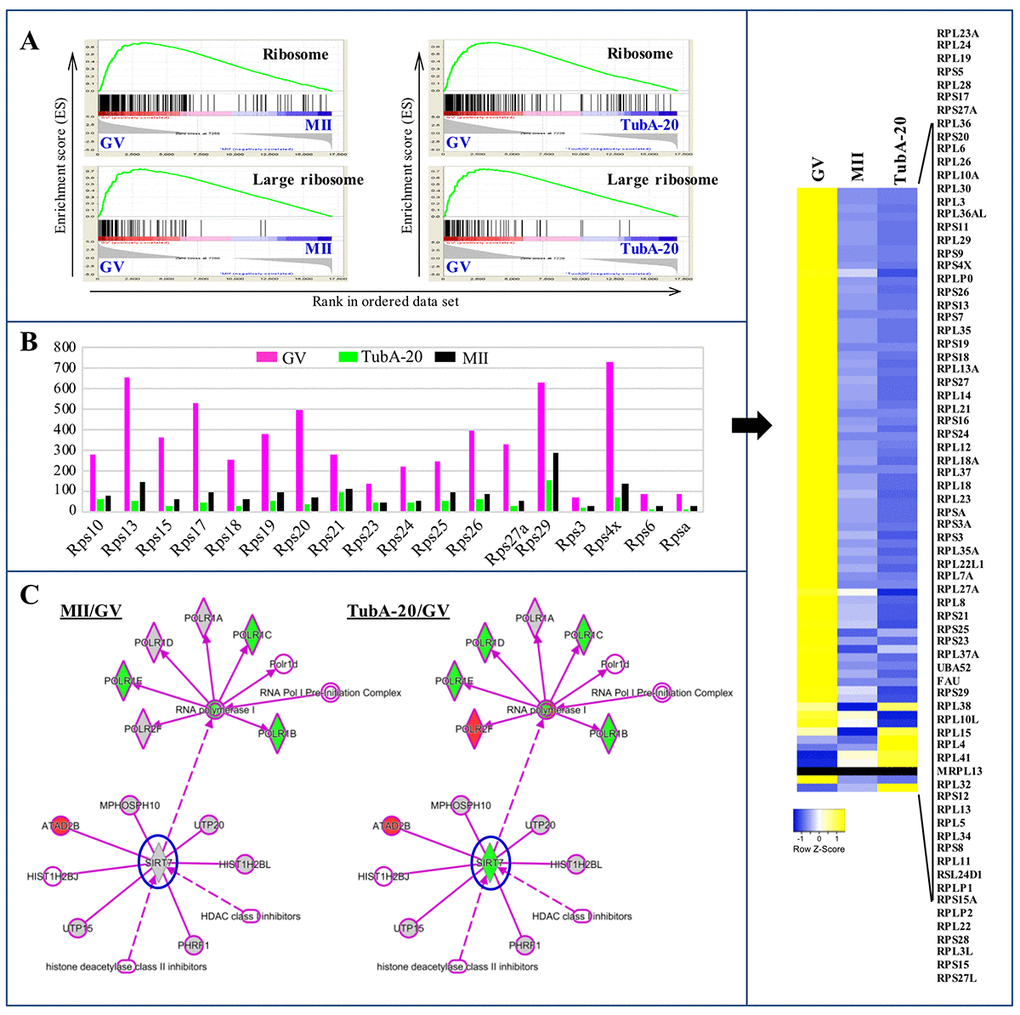
Figure 5.Ribosome biosynthesis in GV, MII, and TubA-treated oocytes. (A) GSEA plots showing preferential downregulation of the KEGG ribosome in matched pairs of TubA20 treated (TubA/GV pairs) versus untreated control (MII/GV pairs). (B) Expression of ribosome biosynthesis-related functional genes in GV-, TubA-20 treated, and MII-stage oocytes. Y-axis shows the FPKM values of genes inferred from the transcriptome data. Right indicates a heatmap showing DEGs of Rps family genes in GV, MII, and TubA-treated oocytes. (C) IPA analysis of MII/GV and TubA/GV pairs. Shapes and lines are color-coded based on predicted associations and functions. Green and red indicate up- and downregulated gene expression.