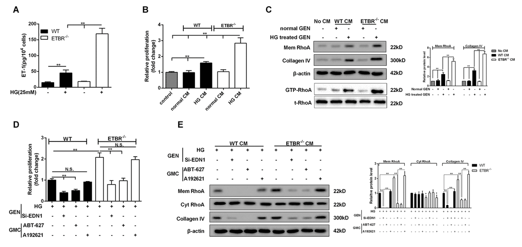
Figure 3.HG conditioned media (CM) of ETBR-/- GENs promoted mesangial cell proliferation and ECM formation. (A) After 24 h of HG (25mM) treatment, ET-1 level in primary GENs of ETBR-/- mice and WT mice was detected by ELISA. CM was collected for the culture of SV40 MES13 cells. **p<0.01 compared with control or HG WT. N=3. (B-C) WT or ETBR-/- CM GEN was used to cultivate SV40 MES13 cells for 24 h. SV40 MES13 cells in control group was cultured in HG serum-free medium. The proliferation of SV40 MES13 cells was detected in control, WT normal CM, WT HG CM, ETBR-/- normal CM and ETBR-/- HG CM groups by MTT assay. RhoA level on SV40 MES13 cells membrane and Collagen IV secretionwere detected in control, WT normal CM, WT HG CM, ETBR-/- normal CM and ETBR-/- HG CM groups by western blot. GTP-RhoA level (the activity of Rho) was detected using Rhotekin RBD-agrose by Rho-pull down assay. **p<0.01 compared with control or normal WT CM or HG WT CM or normal ETBR-/- CM. N=3. (D-E) GENs were transfected with 50 nM si-EDN1 for 18 h, and HG medium was used to culture GENs for 24 h, then the CM was collected for the culture of mesangial cells. 25 μM ABT-627 (blocking agent of ET-1/ETAR pathway) or 25μM A192621 (blocking agent of ET-1/ETBR pathway) was added to the medium for the culture of mesangial cells. The proliferation of mesangial cells was detected by MTT assay, and mem RhoA, cyt RhoA, collagen IV protein levels were detected by western blot. **p<0.01 compared with si-ET-1 or si-ET-1+ABT-627 or si-ET-1+A192621. Bars depict the mean ± SD. N=3.