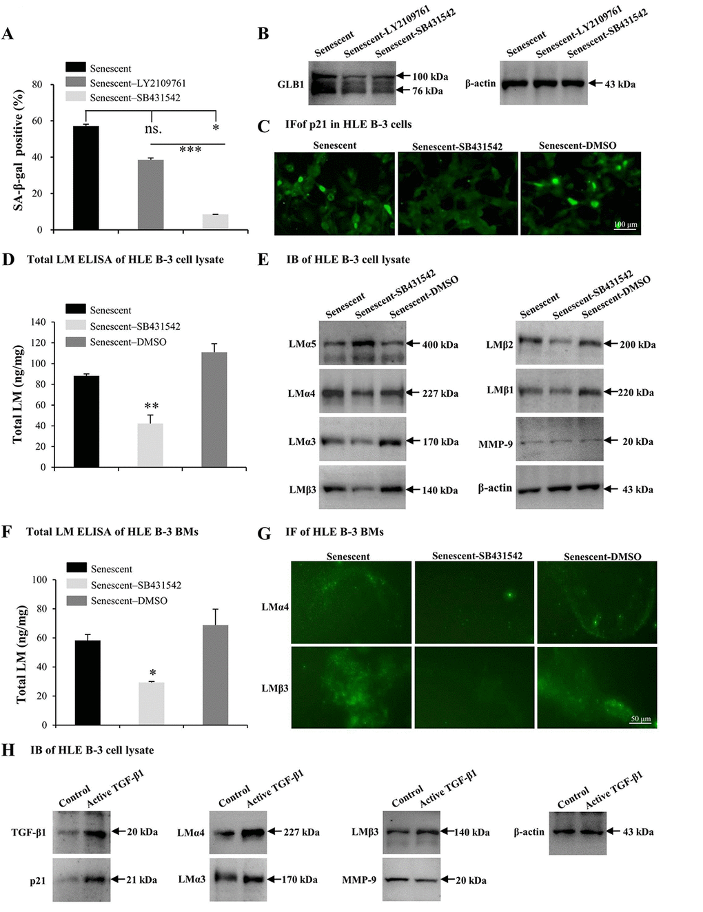
Figure 6.TGF-β1 enhances cell senescence and laminin (LM) deposition induced by H2O2. Senescent HLE B-3 cells [or cell basement membranes (BMs)] were cultured in medium with H2O2 (400 μM) for 96 h. (A-G) HLE B-3 cells were treated with H2O2 only, or in combination with LY2109761 (5 µM) or SB431542 (10 µM) for 72 h. (A-C) Percentage of SA-β-gal-positive cells (A), protein expression of GLB1 (B) and immunofluorescence analysis of p21 (C) in HLE B-3 cells. (D) Total LM in HLE B-3 cells, as detected by ELISA. (E) Immunoblot analysis of LM subunits and MMP-9 in HLE B-3 cells. (F) Total LM in HLE B-3 cell BM, as detected by ELISA. (G) Immunofluorescence analysis of LMα4 (green) and LMβ3 (green) in HLE B-3 cell BMs (Scale bars: 100 μm). (H) HLE B-3 cells treated with TGF-β1 (15 ng/ml) for 96 h. Protein expression levels of TGF-β1, p21, MMP-9 and LM subunits in HLE B-3 cells analyzed via IB. Data were shown as mean ± SD and were analyzed using paired t-test. *, p<0.05; **, p<0.01.