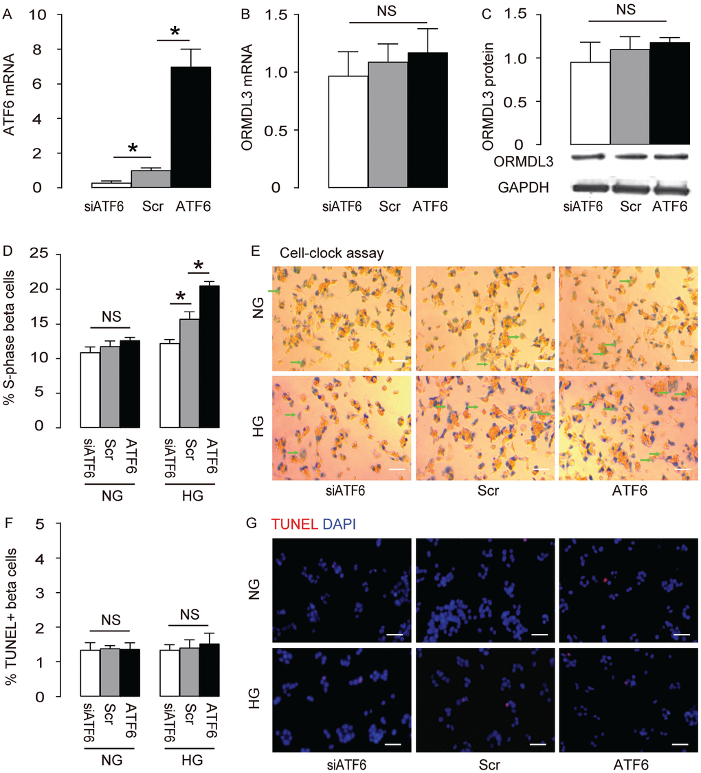
Figure 3.ATF6 increases beta cell proliferation cultured in HG. Min6 cells were kept in high glucose (20mmol/l) culture, and transfected with ATF6, or scrambled (Scr), or siRNA for ATF6 (siATF6). (A–B) RT-qPCR for ATF6 (A) and ORMDL3 (B). (C) Western blot for ORMDL3. (D–E) Cell-clock cell cycle assay, shown by quantification of S-phase cells (D), and by representative images (E). (F–G) TUNEL assay, shown by quantification (F), and by representative images (G). DAPI: nuclear staining. NG: normal glucose culture. HG: high glucose culture. Arrows pointed to S-phase cells. *p<0.05. NS, no significance. N=5. Scale bars are 20μm.