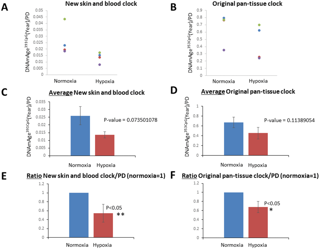
Figure 4.Hypoxia slowed the speed of the cell division-associated DNAm age progression. (A and B) DNAmAge391CpG/PD (A) and DNAmAge353CpG/PD (B) of each cell line in normoxia and hypoxia are shown. The result from the same cell line is marked with the same color. In all cell lines examined, hypoxia slowed the speed of DNAm age progression. (C and D) Average and S.E. of DNAmAge391CpG/PD and DNAmAge353CpG/PD are shown. (E and F) In these graphs, DNAm age/PD in normoxia is designated as 1 in all cell lines, and the ratio of DNAm age in normoxia and hypoxia was calculated. In both DNAmAge391CpG and DNAmAge353CpG, hypoxia slowed the speed of the progression of DNAm age and the effects were statistically significant (t-test).