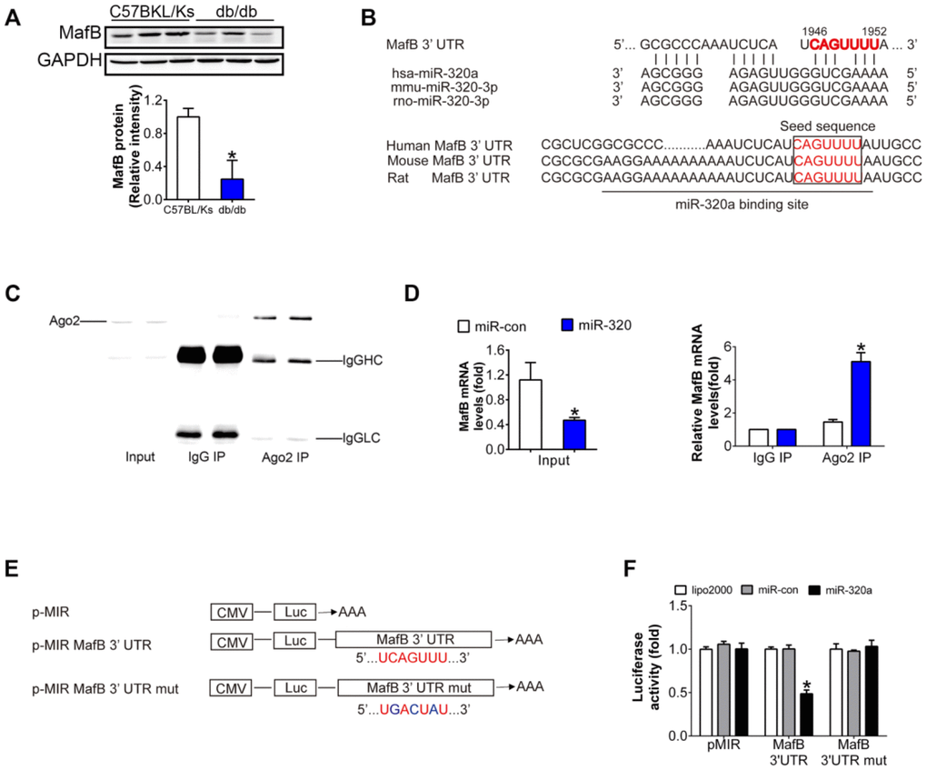
Figure 4.MafB is a target of miR-320a. (A) MafB protein levels detected by western blot in C57BLKS and db/db mice. (B) miR-320a and the 3’-UTR of MafB among three species. (C) Ago2 protein levels in co-immunoprecipitated products detected by Western blot. IgGHC, IgG heavy chain; IgGLC, IgG light chain. (D) Relative expression of MafB in the whole RNA (left) and RNA of the nonspecific IgG or anti-Ago2 co-IP (right) from the HG-treated podocyte cell lysates. #P<0.05 versus miR-con + input, *P<0.05 versus miR-con + IgG IP. (E) Schematic diagram of the luciferase reporter plasmids of pMIR-MafB 3’-UTR and pMIR-MafB 3’-UTR mut, and the potential target site of miR-320a on the 3’-UTR of MafB. (F) Regulation of miR-320a on 3’-UTR of MafB in HEK293 cells by luciferase reporter assay. *P<0.05 versus MafB 3’-UTR + miR-con.