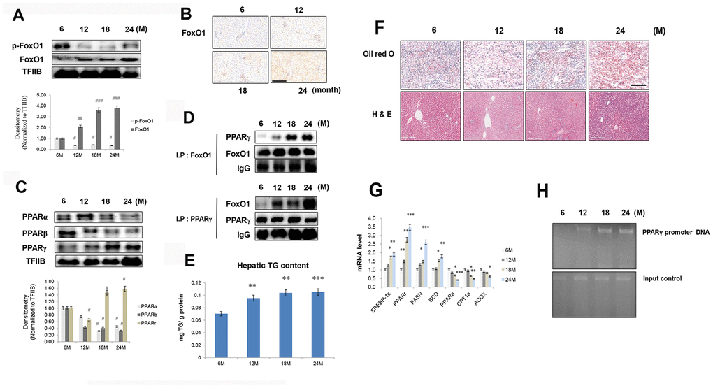
Figure 3.Aging-related increase in FoxO1-induced lipid accumulation. (A) Western blotting was performed to examine the protein levels of p-FoxO1 and FoxO1 in the liver of aging rats. Results of one-factor ANOVA: ##p < 0.01, and ###p < 0.001 vs. 6 months. (B) Immunohistochemical staining for FoxO1 in aging liver. Scale bar: 200 μm. (C) Western blotting analysis of PPARs in the nuclear of aging liver. TFIIB was the loading control of the nuclear fraction. One representative result of the three experiments for each protein is shown. Results of one-factor ANOVA: #p < 0.05 vs. 6 months. (D) Western blotting showed that immunoprecipitated FoxO1 and PPARγ were physically associated with PPARγ and FoxO1, respectively. (E) Hepatic TGs in aging rats. Results of one-factor ANOVA **p < 0.01, and ***p < 0.001 vs. 6 months. (F) Aging livers were stained with Oil red O to visualize lipid accumulation. Scale bar: 100 μm. Representative H&E staining shows increased vacuoles in liver tubules during aging. Scale bar: 300 μm. (G) Real-time PCR analyses was performed for measuring the mRNA levels of SREBP-1c, PPARγ, FASN, SCD, PPARα, CPT1α, and ACOX. The data are expressed as a mean ± SEM. *p < 0.05, **p < 0.01, and ***p<0.001 vs. 6 months. (H) FoxO1 binds to the PPARγ promoter in aging livers. The livers were subjected to ChIP assay by using rabbit pre-immune IgG and an anti-FoxO1 antibody. Immunoprecipitates were subjected to PCR by using rat PPARγ promoter DNA.