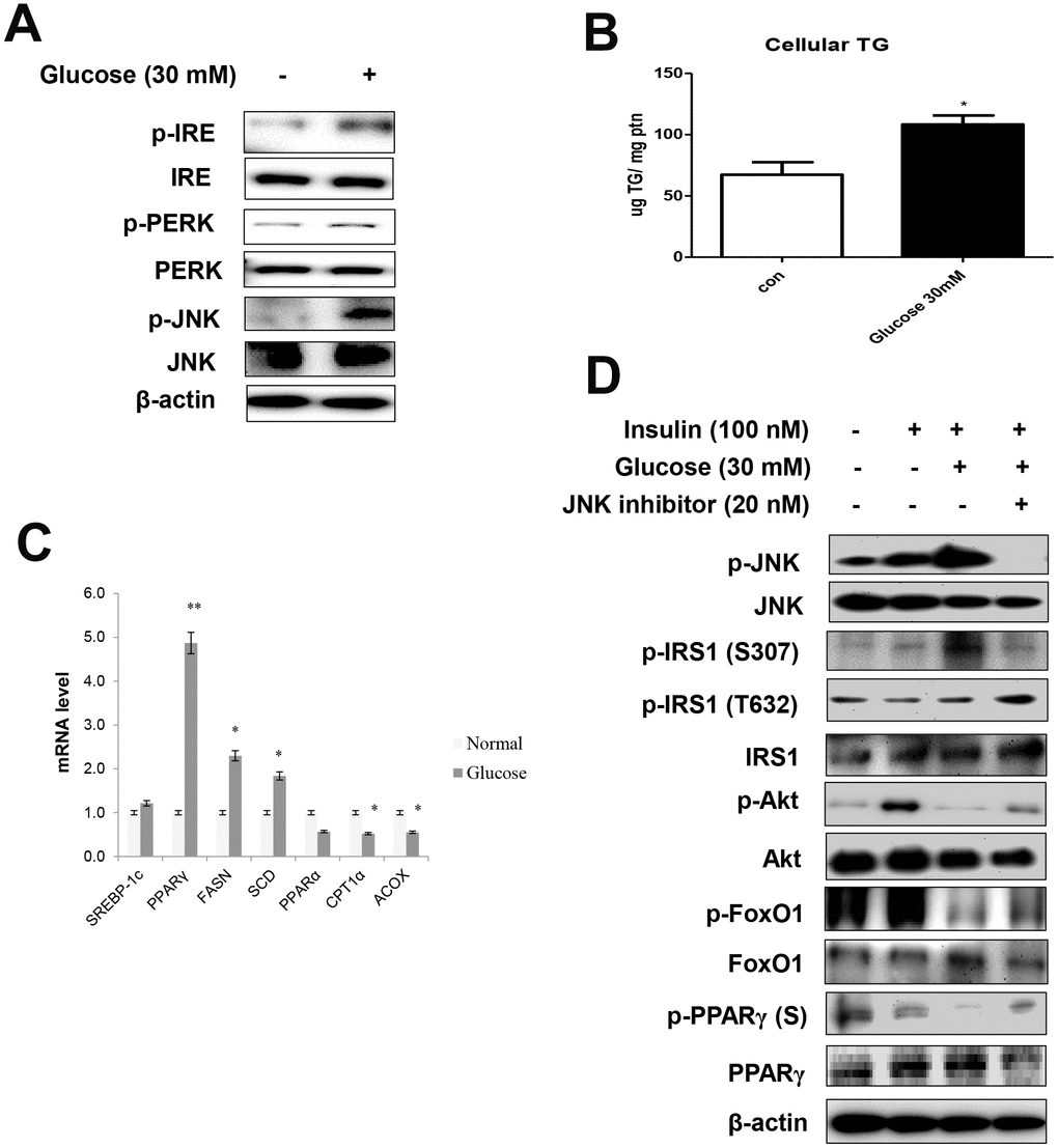
Figure 4.High glucose induced ER stress-mediated lipid accumulation. (A) Western blot was used to detect p-IRE, total-IRE, p-PERK, total-PERK, p-JNK, and total-JNK in cytoplasmic extracts (20 μg protein) after treatment of AC2F cells with glucose (30 mM) for 6 h. β-actin was the loading control of the cytosolic fractions. (B) Cellular triglyceride concentration after treatment with glucose (30 mM) for 36 h was measured by a colorimetric assay. The data are expressed as a mean ± SEM. Three independent experiments were performed and similar results were obtained. *p < 0.05 vs. non-treated cells. (C) Real-time PCR analyses was performed for measuring the mRNA levels of lipogenesis genes (SREBP-1c, PPARγ, FASN, SCD) and β-oxidation genes (PPARα, CPT1α, and ACOX). The data are expressed as a mean ± SEM. Three independent experiments were performed and similar results were obtained. *p < 0.05, and **p < 0.01 vs. non-treated cells. (D) After stimulation with glucose (30 mM) for 2 h with insulin (100 nM) for 10 min in the absence (-) or presence (+) of JNK inhibitor (PD98059, 20 μM) for 1 h, cells were lysed and analyzed by western blotting. β-actin was the loading control of the cytosolic fractions.