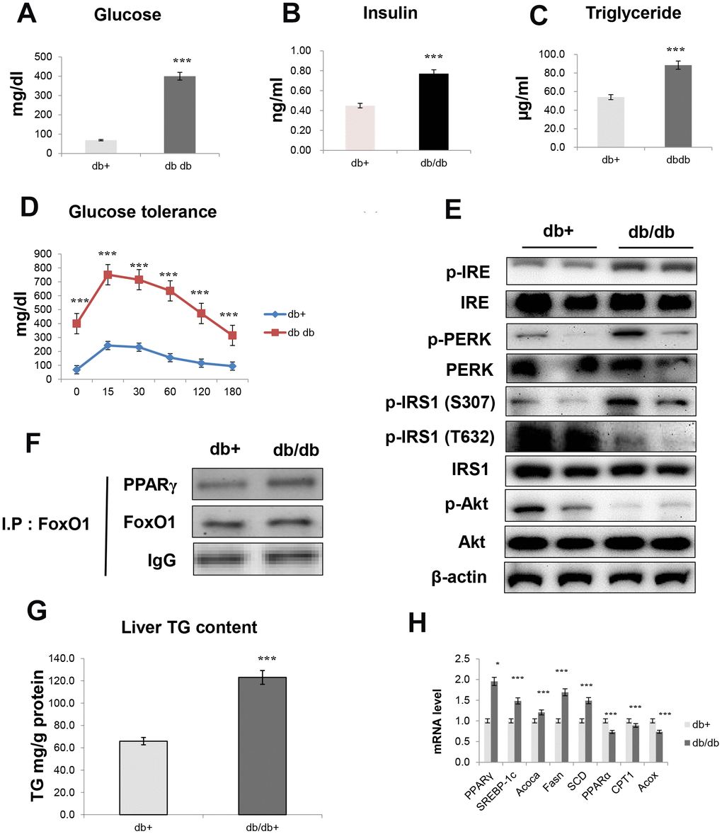
Figure 8.Obesity induces hepatic steatosis and insulin resistance through ER signaling. (A) Glucose level (B) insulin level (C) triglyceride level (D) glucose tolerance test in the serum of obese models. The data are expressed as a mean ± SEM (each n=5). The data are expressed as a mean ± SEM. ***p<0.001 vs. db+. Western blot analyses of liver cytosolic (E) p-IRE, IRE, p-PERK, PERK, p-IRS1 (S307), p-IRS1 (T632), IRS1, p-Akt, and total-Akt levels were performed using cytosolic proteins from obese mice (n = 5 in each group). β-actin was the loading control of the cytosolic fractions. (F) Western blotting showed that immunoprecipitated FoxO1 and PPARγ were physically associated with PPARγ and FoxO1, respectively. (G) Hepatic triglyceride concentration was measured by a colorimetric assay. The data are expressed as a mean ± SEM. ***p<0.001 vs. db+. (H) PPARγ, SREBP-1c, Acoca, Fasn, SCD, PPARα, CPT1, and Acox mRNA. Real-time PCR analyses were performed to determine the mRNA levels in liver tissues of db/db mice (n = 5 in each group). The data are expressed as a mean ± SEM. *p < 0.05, and ***p<0.001 vs. db+.