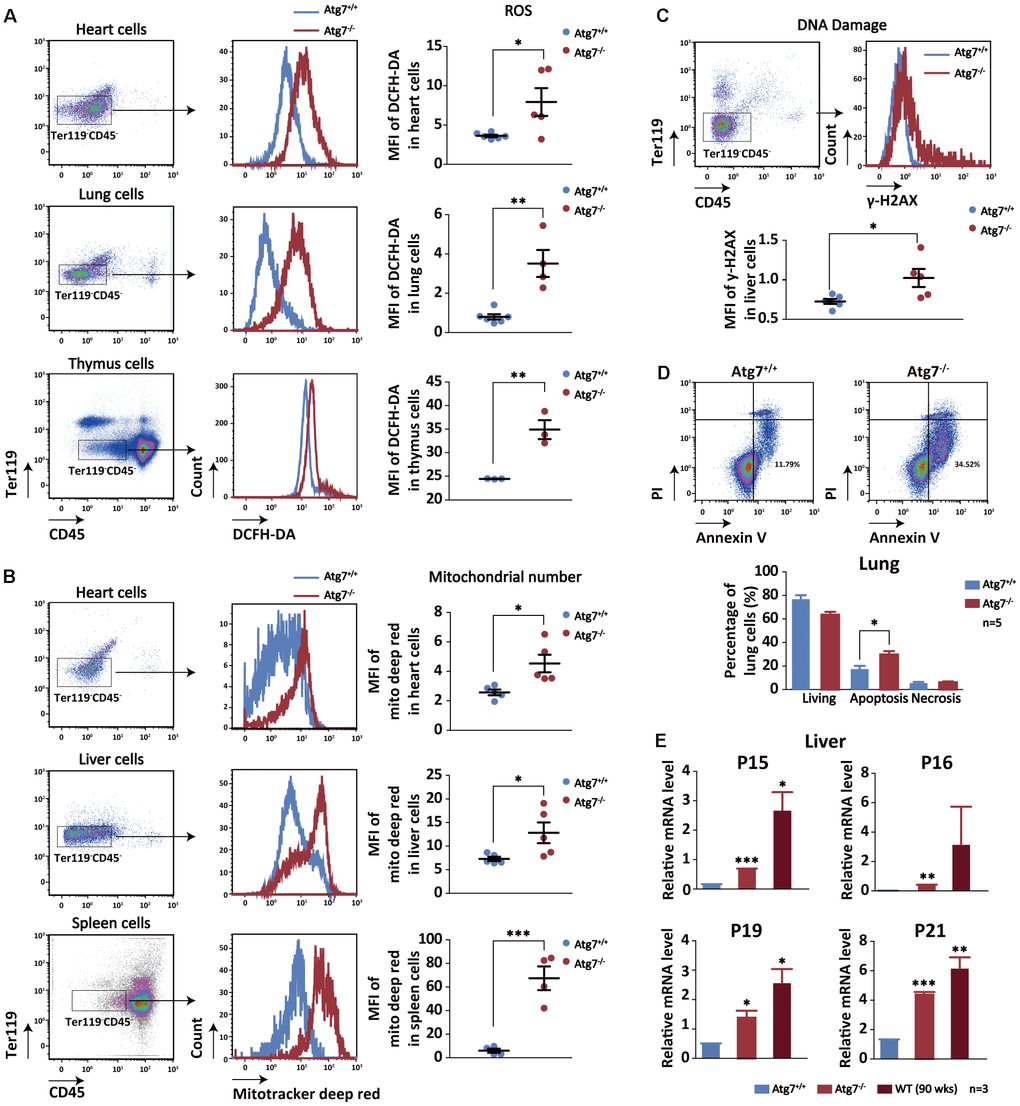
Figure 4.Increased cellular aging markers in the non-hematopoietic organs of the mice with hematopoietic autophagy defect. (A) Flow cytometric analysis of ROS levels of the heart, lung and thymus cells with fluorescein DCFH-DA. Left and middle, gating strategy for the flow-cytometric assessment of non-hematopoietic cells (CD45-Ter119-); right, geometric mean fluorescence intensity (MFI) of DCFH-DA in the heart cells of wild-type mice and Atg7-deleted mice. (B) Flow cytometric analysis of mitochondrial mass levels of the heart, liver and spleen cells with florescent Mitotracker Deep Red. Left and middle, gating strategy for the flow-cytometric assessment of non-hematopoietic cells (CD45-Ter119-); right, geometric mean fluorescence intensity (MFI) of MitoTracker Deep Red in the heart or liver cells of wild-type and Atg7-deleted mice. (C) Flow cytometric analysis of DNA damage with γ-H2AX. Upper, gating strategy for non-hematopoietic cells (CD45-Ter119-) in the liver; lower, geometric mean fluorescence intensity (MFI) of γ-H2AX in the liver cells. (D) Analysis of apoptosis and necrosis in the lung cells of wild-type mice and Atg7-deleted mice by annexinV and PI double staining. Upper, representative flow cytometric measurement; lower, statistical results from cytometric analysis. (E) Quantitative RT-PCR analysis of four aging related genes (P15, P16, P19, P21) in the liver cells (blood cells removed by sorting against CD45+ or Ter119+) of young wild-type mice, Atg7-deleted mice and old wild-type mice.