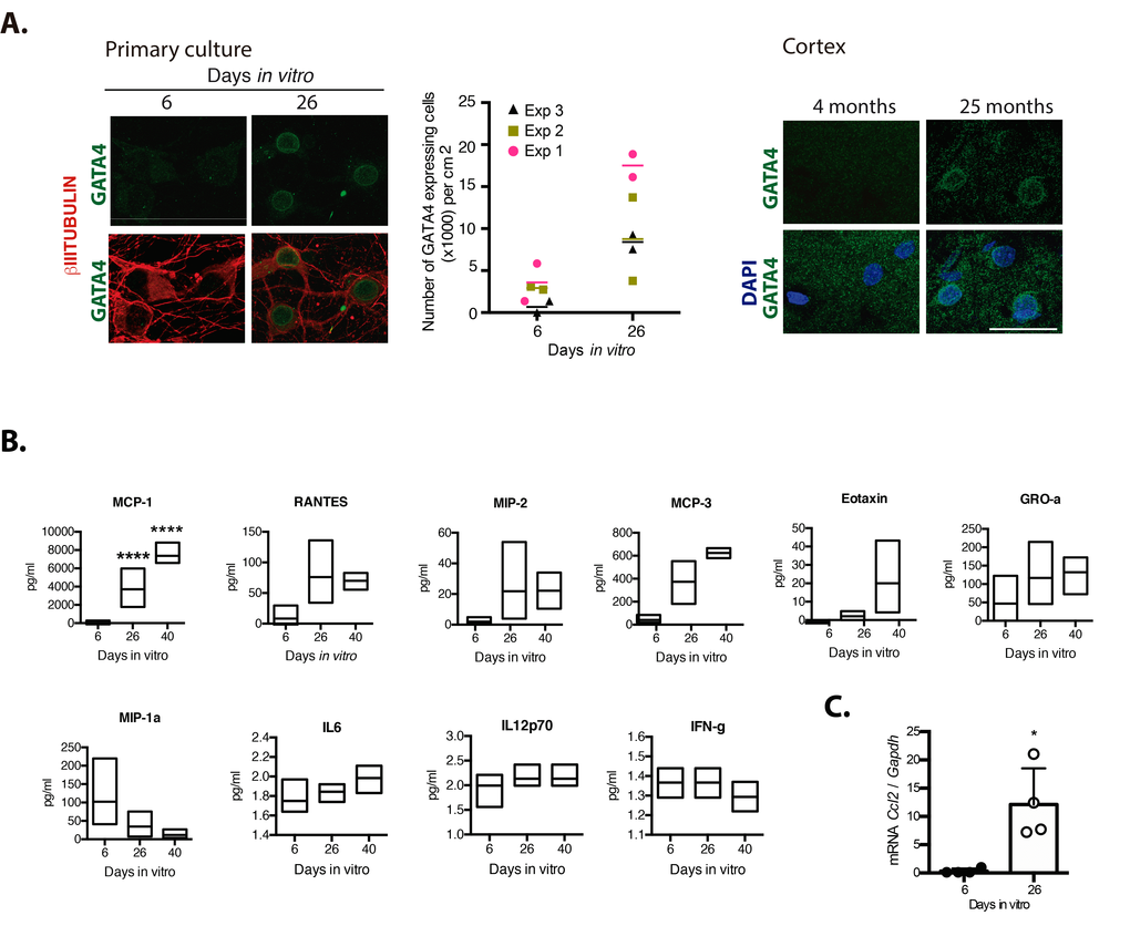
Figure 5.Senescent neurons increased the expression of GATA4 and cortical cells secreted MCP-1. (A) Immunofluorescence to detect GATA4 in neurons (expressing βIII-TUBULIN) in primary culture of cortical cells either incubated during the indicated days in vitro or in rat brains of the indicated age. Scale bars represent 25 μm. The number of cells with increased GATA4 abundance from three independent experiments, each performed in duplicate, is graphed. The mean of each experiment is represented by horizontal bars. (B) Quantification by multiplex immunoassay of the indicated cytokines, from conditioned media from cultures of the indicated days from three independent experiments. The maximum and minimum values are graphed. Bars indicate the mean of the three independent experiments. Data were analyzed by two-way ANOVA followed by Tukey´s multiple comparisons test analysis, only MCP-1 was significant. **** p<0.0001 relative to 6 DIV. (C) qRT-PCR from total RNA purified from cortical primary cultures during the indicated days. The relative expression of Ccl2 mRNA was normalized with Gapdh mRNA. Bars represent SD. * p=0.0106 by unpaired t test two tailed. n=4.