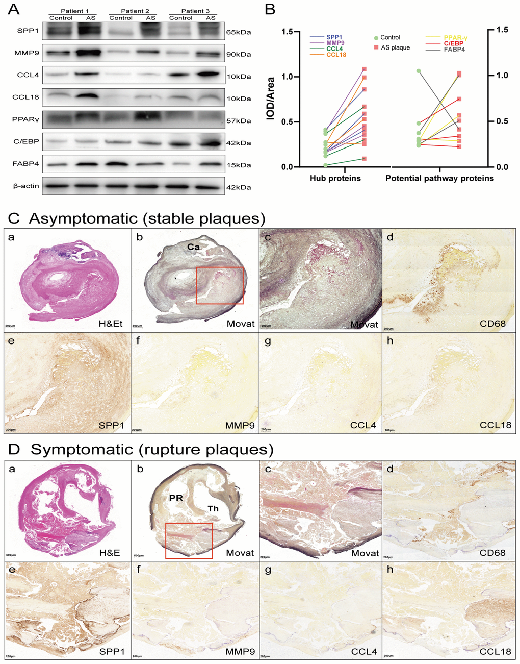
Figure 7.Hub proteins and potential pathway proteins were increased in AS plaques, especially in tissue sections of ruptured plaques. (A) Detection of hub proteins (SPP1, MMP9, CCL4, and CCL18) and potential pathway proteins (PPARγ, C/EBP and FABP4) in ruptured plaques and adjacent normal tissues by western blots. β-actin was used as a loading control. Bands were quantified with ImageJ software. (B) Line chart showing IOD/area of proteins in immunoblot analysis. Red squares represent AS plaques, and the green circles indicate adjacent normal tissues. The different colored lines represent the trend of protein expression. (C, D) H&E (a) and Movat (b and c, low and high power) staining and immunoperoxidase antibody staining using anti-CD68 (d), anti-SPP1 (e), anti-MMP9 (f), anti-CCL4 (g), and anti-CCL18 (h). Ca, calcification; PR, plaque rupture; Th, thrombus.