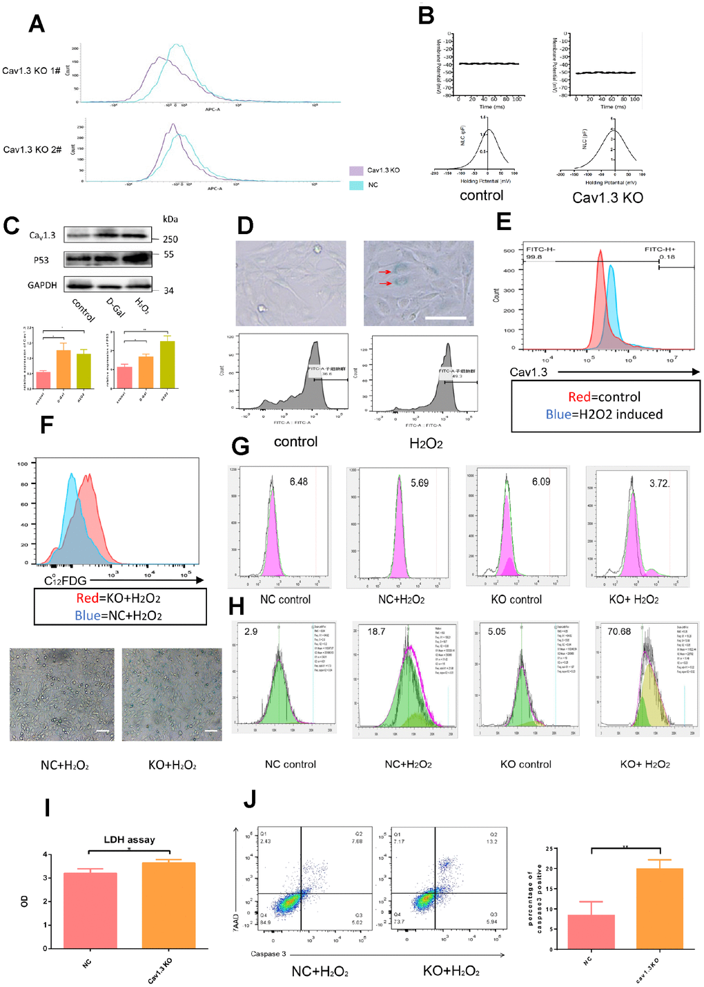
Figure 3.Hair cells were vulnerable to ROS injury after Cav1.3 was knocked out. (A) the effect of CaV1.3 knock out in HEI-OC1 was analyzed by flow cytometry. (B) membrane potential (top) and non-linear capacitance (NLC) (bottom) studies in WT HEI-OC1 and CaV1.3 KO HEI-OC1 cells (n=5). (C) western-blotting analysis of CaV1.3 and p53 expression in control and senescence HEI-OC1 cells induced by D-galactose (D-Gal) or hydrogen peroxide (H2O2), the bottom panel is the quantitative analysis. (D) β-Galactosidase staining (top) and C12FDG staining (bottom) of control and senescent HEI-OC1 cells induced by H2O2. (E) flow cytometry analysis of CaV1.3 in control and H2O2 induced HEI-OC1 cells. (F) C12FDG staining (top) and β-Galactosidase staining (bottom) of NC (negative control) and KO (CaV1.3 knock out) HEI-OC1 cells after H2O2 induction. (G, H) CFSE staining and red dot staining of NC and KO HEI-OC1 cells with or without H2O2 induction. (I) LDH assay of NC and KO HEI-OC1 cells after H2O2 induction (n=3). (J) caspase-3/7-AAD staining of NC and KO HEI-OC1 cells after H2O2 induction (n=3). Error bars represent mean ± s.d.; *P<0.05; **P < 0.01; ***P < 0.001; n.s. not significant; by one-way analysis of variance (ANOVA) (I, J).