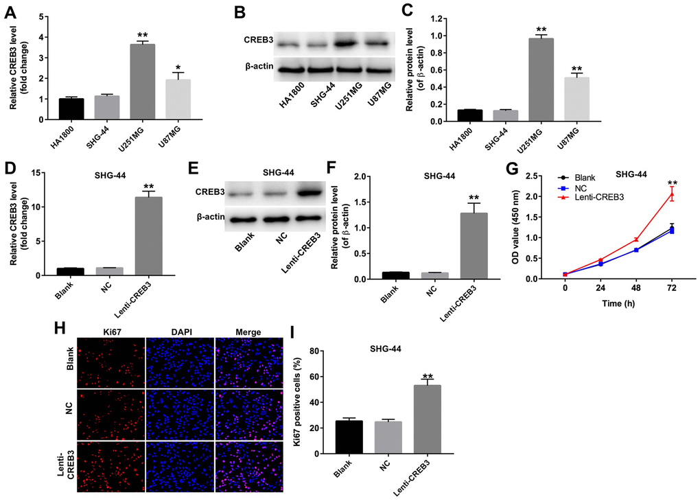
Figure 3.The upregulation of CREB3 promoted the proliferation of SHG-44 cells. (A) The relative levels of CREB3 in four cell lines (HA1800, SHG-44, U251MG and U87MG cells) were detected by qRT-PCR. (B, C) The relative levels of CREB3 in HA1800, SHG-44, U251MG and U87MG cells were detected by Western blotting. β-actin was used as a loading control. (D) CREB3 levels in SHG-44 cells transfected with the NC or lenti-CREB3 were detected by qRT-PCR. (E, F) CREB3 levels in SHG-44 cells transfected with the NC or lenti-CREB3 were measured by Western blotting. β-actin was used as a loading control. (G) The viability of SHG-44 cells transfected with the NC or lenti-CREB3 was detected with a CCK-8 assay at 0, 24, 48 and 72 h. (H, I) The relative fluorescence levels were quantified for KI67 and DAPI staining. *P<0.05, **P<0.01 compared with the HA1800 group or NC group.