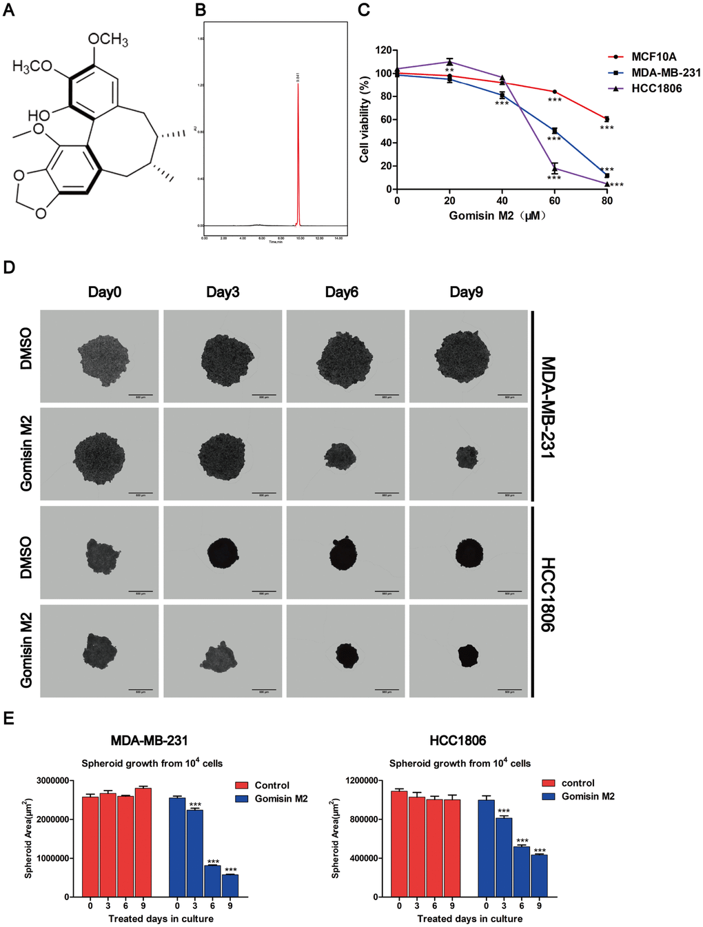
Figure 1.Effects of Gomisin M2 on the viability of MCF10A, MDA-MB-231, and HCC1806 cells. (A) The chemical structure of Gomisin M2. (B) The HPLC chromatograms of Gomisin M2. (C) Cells were treated with increasing doses of Gomisin M2 for 48 h. Cell viability determined by Alamar blue assay. (D) Images of the 3D spheroids that were treated with Gomisin M2 over 9 days were acquired in all microplates using the PerkinElmer Operetta High-Content Imaging System. Scale bar = 200 μm. (E) Bar plot of the average cross-sectional area of the MDA-MB-231 and HCC1806 spheroids. Approximately three replicate tumor spheroid samples were used for quantification. The data were expressed as the mean ± SD. Compared with the DMSO group: **p < 0.01.