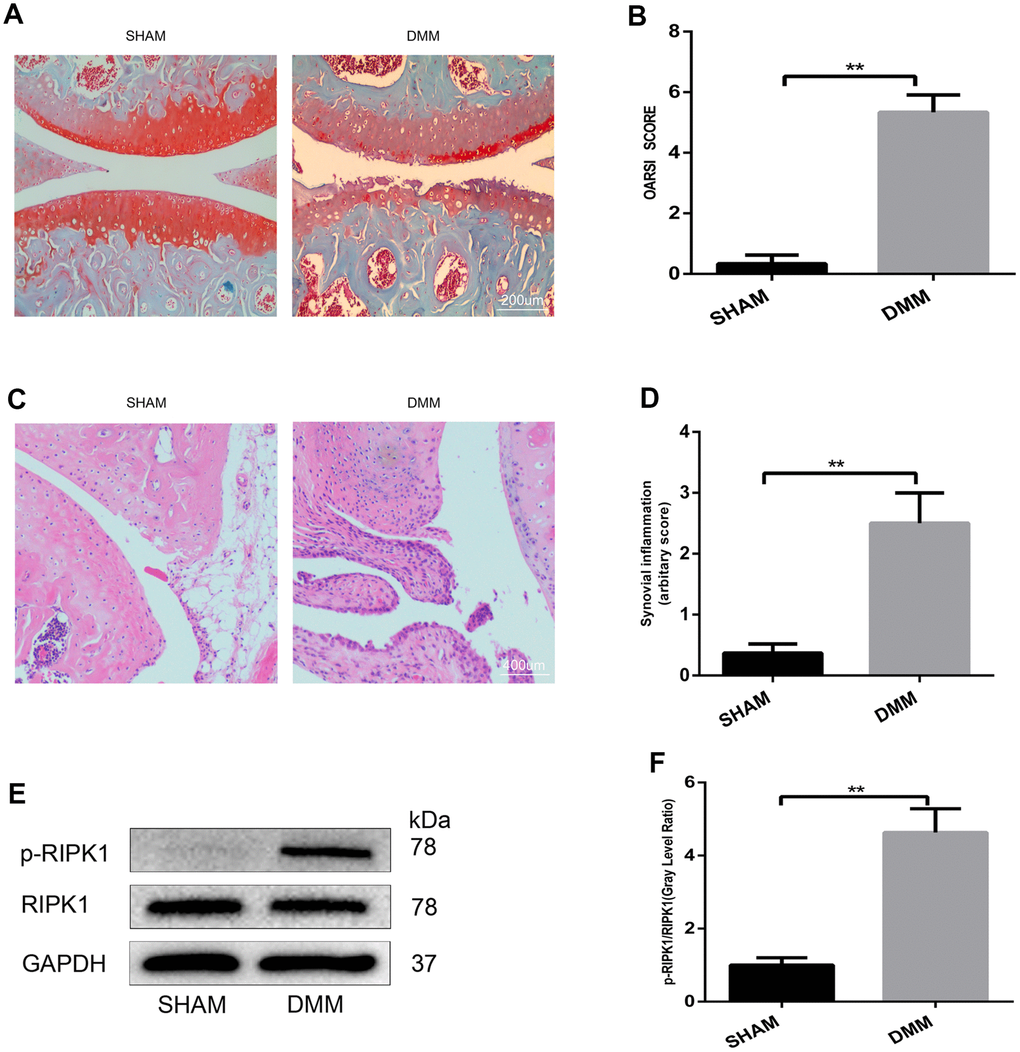
Figure 1.The phosphorylation level of RIPK1 is increased in mouse knee articular cartilage. (A, B) Safranin O/Fast Green-stained sagittal-plane images of tibial and femoral cartilage from the sham and destabilized medial meniscus (DMM) groups; the Osteoarthritis Research Society International (OARSI) score was significantly increased in the DMM group (n = 10); scale bar = 200 μm. (C, D) Representative hematoxylin and eosin (HE)-stained images and synovial inflammation scores in the sham and DMM groups (n = 10); scale bar = 400 μm. (E, F) Western blots and quantitative data of p-RIPK1 in the sham and DMM groups. The experiments were repeated three times independently. Columns represent means ± SD. **p < 0.01.