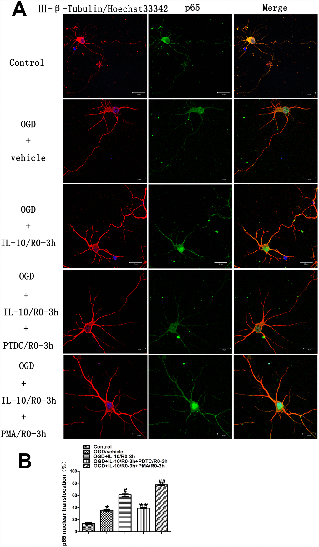
Figure 4.Localization and nuclear translocation of p65 at an early stage after OGD injury. (A) Representative images of each group. The left column displays neuronal marker class Ⅲ-β-Tubulin (red) and nucleus (blue). The middle column shows expression of p65 (green). The right column indicates the co-localization of class Ⅲ-β-Tubulin, nucleus and P65. Scale bar is 20 μm. (B) Quantification of p65 nuclear translocation. *p<0.001, as compared with Control group; #p<0.001, as compared with OGD group; **p<0.001, as compared with OGD+IL-10/R0-3h group; ##p<0.001, as compared with OGD+IL-10/R0-3h group; by one way analysis of variance (ANOVA) followed by Student-Newman-Keuls multiple comparison test, F=225.278, p<0.0001. All data are presented as mean±SEM.