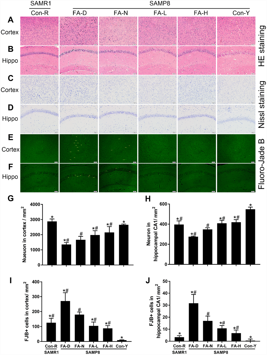
Figure 3.FA supplementation delayed neurodegeneration and stimulated neuronal survival in cerebral cortex and hippocampal CA1 region in SAMP8 mice. Mice were assigned to treatment groups as described in Figure 1. Representative micrographs of HE staining in cerebral cortex (A) and hippocampal CA1 region (B), Nissl staining in cerebral cortex (C) and hippocampal CA1 region (D), Fluoro-Jade B (FJB) staining in cerebral cortex (E) and hippocampal CA1 region (F). Quantitative analysis of surviving neurons indicated by Nissl staining in cerebral cortex [F(5,24) = 30.882, P<0.001] (G) and hippocampal CA1 region [F(5,24) = 68.256, P<0.001] (H), and degenerated neurons indicated by FJB-positive cells in cerebral cortex [F(5,24) = 44.797, P<0.001] (I) and hippocampal CA1 region [F(5,24) = 45.630, P<0.001] (J). Yellow arrow indicates FJB positive cell. Scale bar = 50 μm. Data are expressed as mean ± SD (n= 5 mice/group). *P<0.05 compared with FA-N group. #P<0.05 compared with Con-Y group.