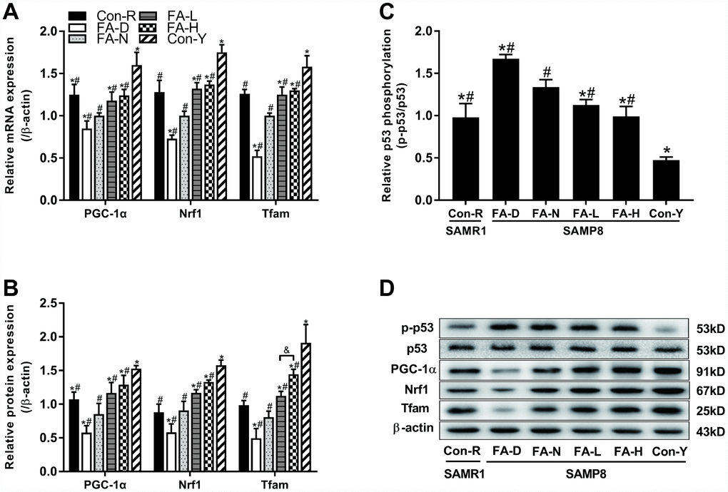
Figure 5.FA supplementation regulated telomere–p53–mitochondria pathway in brain of SAMP8 mice. Mice were assigned to treatment groups as described in Figure 1. (A) The relative mRNA levels of PGC-1α, Nrf1 and Tfam quantified by qPCR were normalized to β-actin and expressed as fold changes relative to the FA-N group [F(5,54) = 30.274, P<0.001; F(5,54) = 36.428, P<0.001; F(5,54) = 34.239, P<0.001]. (B) The protein expressions of PGC-1α, Nrf1 and Tfam quantified by western blot were normalized to β-actin [F(5,12) = 21.689, P<0.001; F(5,12) = 39.452, P<0.001; F(5,12) = 38.230, P<0.001]. (C) The level of phospho-p53 quantified by western blot was normalized to p53 [F(5,12) = 51.308, P<0.001]. (D) Representative western blot of phospho-p53, p53, PGC-1α, Nrf1, Tfam and β-actin. Data are expressed as mean ± SD (n= 10 mice/group for qPCR, and n= 3 mice/group for western blot). *P<0.05 compared with FA-N group. #P<0.05 compared with Con-Y group. &P<0.05 compared between FA-L and FA-H groups.