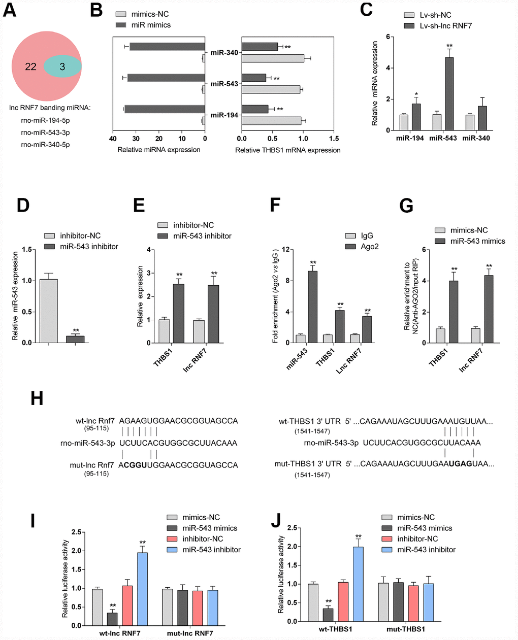
Figure 6.Selection and validation of miRNA that might target lncRNA RNF7 and THBS1. (A) A schematic diagram showing 25 miRNAs that might target THBS1 predicted by online tools; of them, 3 were predicted to target lncRNA RNF7. (B) The overexpression of these 3 miRNAs, miR-340, miR-543, and miR-194, was achieved in rat cardiac fibroblasts by transfection of miRNA mimics, as confirmed by real-time PCR. THBS1 mRNA expression in response to miRNA overexpression was determined by real-time PCR, respectively. (C) Rat cardiac fibroblasts were infected by Lv-sh-lnc RNF7 and examined for the expression of these miRNAs. (D) miR-543 inhibition achieved in rat cardiac fibroblasts by transfection of miR-543 inhibitor and confirmed by real-time PCR. (E) The expression of THBS1 and lnc-RNF7 in response to miR-543 inhibition was determined in cardiac fibroblasts by real-time PCR. (F) The levels of miR-543, THBS1, and lnc RNF7 precipitated by anti-AGO2 antibody were determined using RIP assays. (G) Endogenous THBS1 or lnc RNF7 pull-down by AGO2 upon overexpression of miR-543 was determined using RIP assays. (H) A schematic diagram showing the structures of wild- or mutant-type THBS1 3′-UTR or lnc RNF7 luciferase reporter vectors (wt-THBS1 3′-UTR/lnc RNF7 and mut-THBS1 3′-UTR/lnc RNF7). Mutant-type vectors contained a 4 bp mutation in the predicted miR-543 binding site. (I, J) These vectors were co-transfected into rat cardiac fibroblast with miR-543 mimics/inhibitor and the luciferase activity was determined.