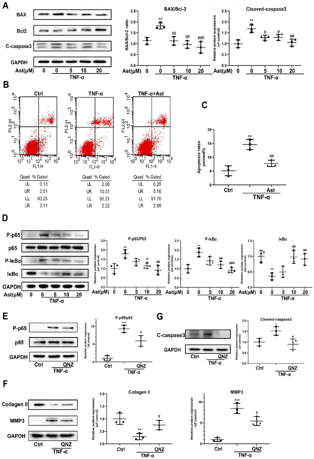
Figure 6.Effects of Ast on TNF-α-induced apoptosis and ECM degradation. (A, B) Protein levels of BAX, Bcl-2, and cleaved caspase-3 were determined by western blotting, and apoptotic chondrocytes were stained with Annexin V-FITC/PI and examined by flow cytometry after treated as above for 24 h. Apoptosis rate was calculated and the data were expressed in (C). FL1 represents Annexin V-FITC and FL2 represents PI. To detect the activation of NF-κB signaling, chondrocytes were serum-starved for 6 h followed by treatment with the vehicle or Ast (5,10, and 20 μM) for 2 h. Cells were then stimulated with TNF-α (5 ng/ml) for 15 min. (D) Activation of the NF-κB signaling pathway was measured by western blotting and quantified. (E) Phosphorylation of p65 was detected by western blotting after the chondrocytes were pre-treated with the vehicle or QNZ (an inhibitor of the NF-κB pathway) for 2 h, followed by stimulation with TNF-α (5 ng/ml). (F, G) Chondrocytes were treated as indicated for 24 h. The expression of Collagen II, MMP3 and cleaved-caspase3 was determined using western blotting and quantified. The data are presented as dot plots from three independent experiments. Significant differences among different groups are indicated as *p<0.05 vs. control; **p<0.01 vs. control; ***p<0.001 vs. control. #p < 0.05 vs. TNF-α group; ##p < 0.01 vs. TNF-α group; ###p < 0.001 vs. TNF-α group.