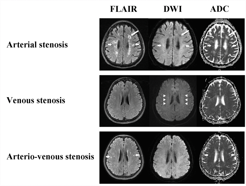
Figure 2.White matter changes of CAS, CVS and CAVS on MRI maps. Arterial stenosis always causes cavity (long arrows) and non-symmetrical multiple round, ovoid, patch and fused hyperintensity lesions with clear boundaries (short arrows). Venous stenosis conduces bilateral and symmetrical cloudy-like white matter hyperintensity surrounding ventricles and centrum semiovale (arrow heads). Arterio-venous stenosis also has non-symmetrical focal white matter lesions (short arrows).