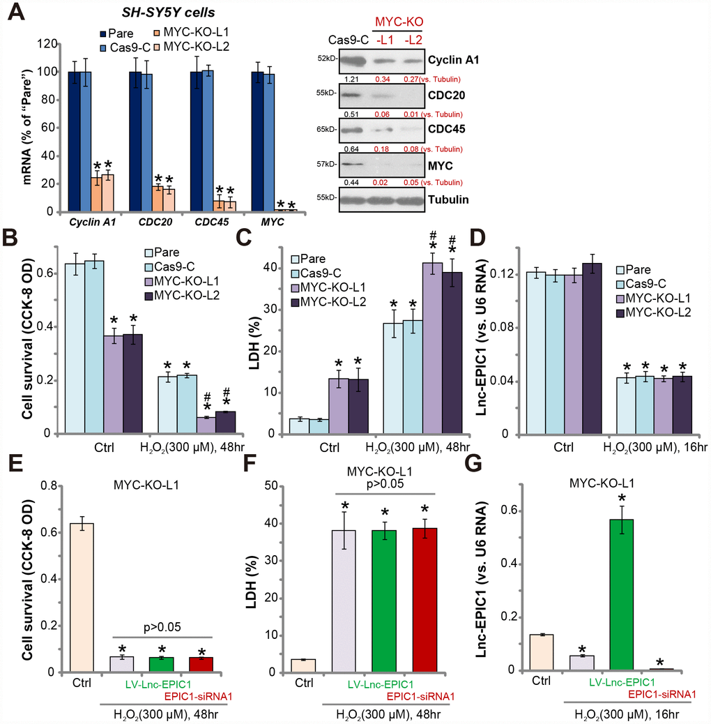
Figure 4.MYC knockout abolishes Lnc-EPIC1-induced actions in H2O2-stimulated neuronal cells. Expression of listed mRNAs and proteins in the stable SH-SY5Y cells with the lenti-CRISPR/Cas9-MYC-KO-GFP constructs (“MYC-KO-L1/L2”) or the control construct (“Cas9-C”) as well as in the parental control cells (“Pare”) were shown (A). Above cells were treated with/without hydrogen peroxide (H2O2, 300 μM) for the applied time, cell viability (by CCK-8 assay, B) and death (by LDH assay, C) were examined; Lnc-EPIC1 levels were tested by qPCR assay (D). “MYC-KO-L1” cells were transfected with lentiviral Lnc-EPIC1 construct (“LV-Lnc-EPIC1”) or Lnc-EPIC1 siRNA-1 (“EPIC1-siRNA1”, 500 nM) for 48h, followed by hydrogen peroxide (H2O2, 300 μM) stimulation for the applied time, cell viability (E), cell death (F) and Lnc-EPIC1 expression (G) were tested. Listed proteins were quantified, with the values normalized to Tubulin (A). Bars stand for mean ± standard deviation (SD, n=5). * P < 0.05 vs. “Ctrl” treatment of “Pare” cells (A–D). * P < 0.05 vs. “Ctrl” treatment (E–G). #P < 0.05 vs. H2O2 treatment of “Pare” cells (A–C). Experiments in this figure were repeated three times, and similar results were obtained.