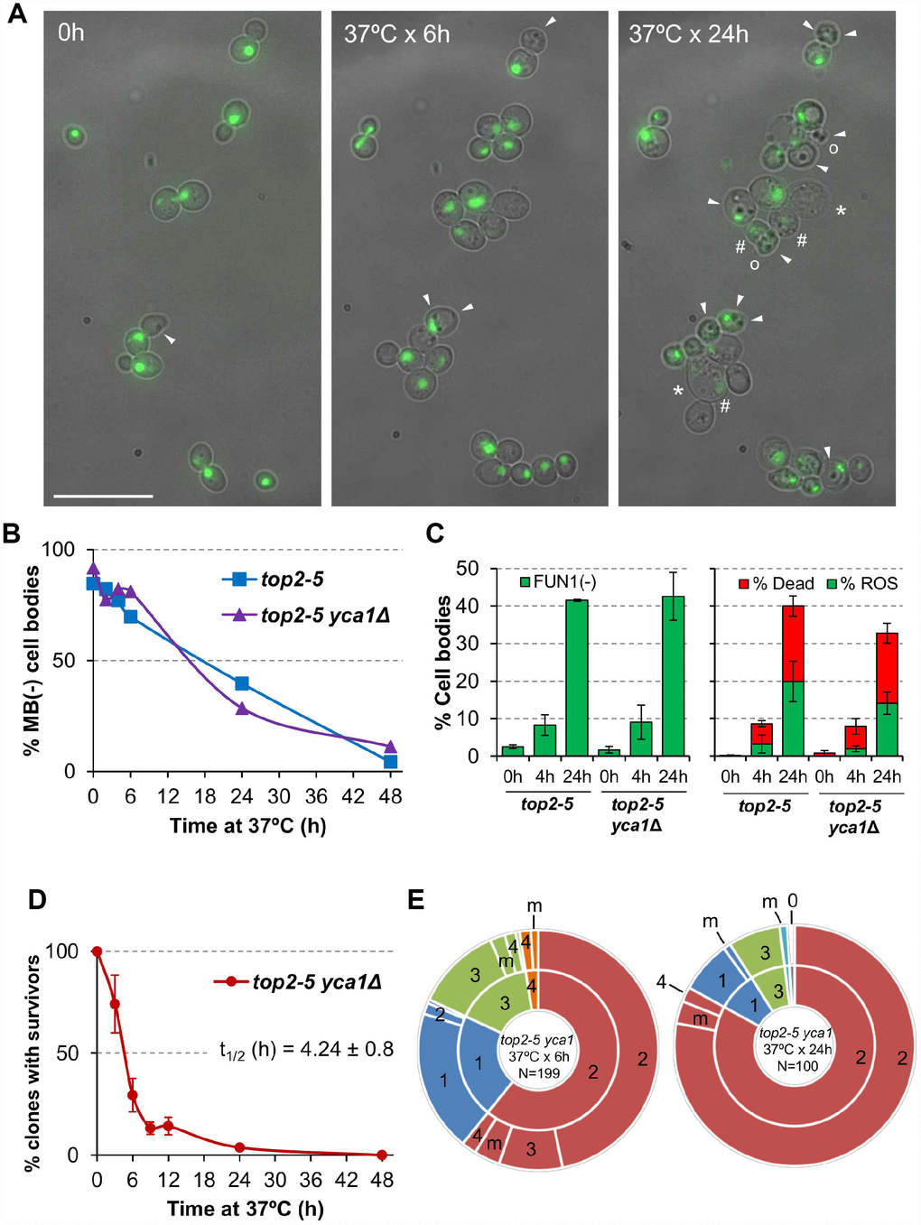
Figure 3.Cell vitality remain high for several hours after the top2 mitotic catastrophe and is not modulated by Yca1. (A) Morphological patterns of cell and nuclear sickness after the top2 MC. Haploid top2-5 HTA2-GFP cells were seeded onto agarose patches and the same fields visualized under the fluorescence microscope at 0 h, 6 h and 24 h after the 37 °C temperature upshift. White filled triangles point to darkened inclusion bodies, asterisks (*) swelled cells, open circles (○) cells that has lost their rounded shape, and hash (#) points to cells that have largely lost the H2A-GFP signal. BF, bright field. Scale bar corresponds to 20 μm. (B) Time course of cell vitality decline as reporter by methylene blue (MB) negative staining. Asynchronous cultures of the top2-5 and top2-5yca1Δ strains were grown at 25 °C before shifting the temperature to 37 °C. At the indicated time points (0, 2, 4, 6, 24 & 48 h), samples were taken and stained with the vital dye MB. (C) Cell vitality decline as reported by metabolic competence, intrinsic ROS generation, and loss of plasma membrane impermeability. Cells were treated as in B and stained at the indicated time points with the vital dye FUN1, the death marker propidium iodide (PI), and/or the ROS reporter DCFH-DA (mean ± s.e.m., n=3). (D) Clonogenic survival profile of top2-5 yca1Δ as determined on the low-density plates (mean ± s.e.m., n=3). The experimental procedure is described in Figure 1D. (E) Ability to re-bud of the top2-5 yca1Δ MC progeny as determined on the high-density plates. The experimental procedure is described in Figure 2.