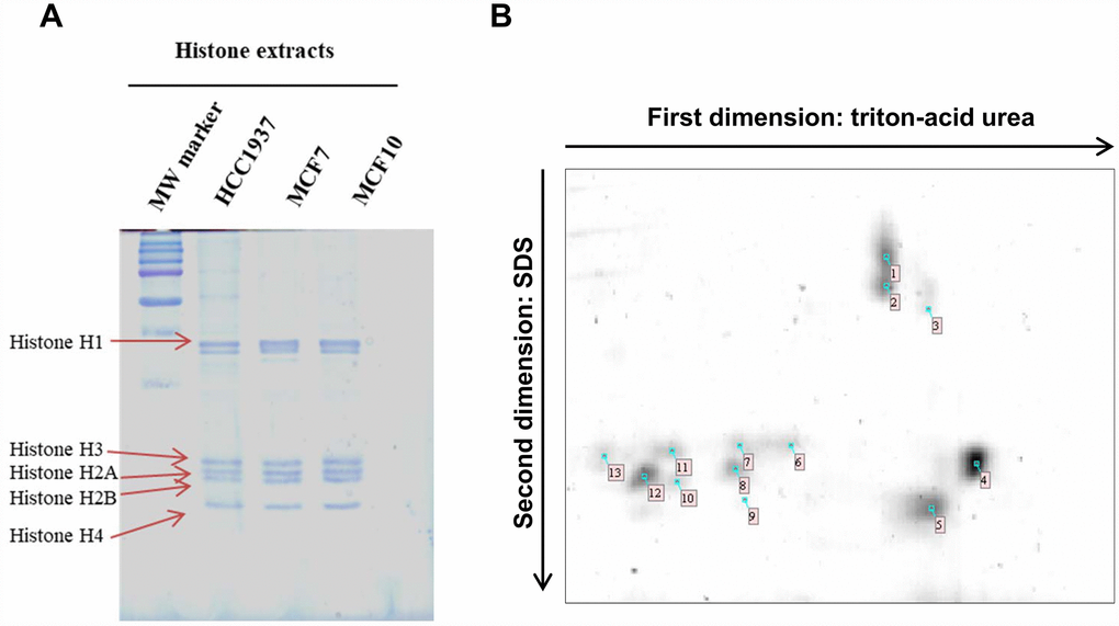
Figure 1.1D TAU gel and 2D TAU gel map of histones in breast cancer cells. (Panel A) The image shows a peculiar separation pattern of histone isoforms, extract from HCC1937, MCF7 and MCF10 cells lines, using 1D-TAU gel. (Panel B) Representative 2D TAU PAGE of histones extract from MCF7 cells. Histones were first resolved by TAU gel and subsequently separated using SDS gel. Spots extracted and analyzed by mass spectrometry are noted on the gel map. All experiments were repeated three times using biologic replicates. Numbered spots are described on table 1 where for each spot is reported the id number, the accession number, histone description, the number of identified peptides, the percentage of sequence coverage, molecular weight and isoelectric point.