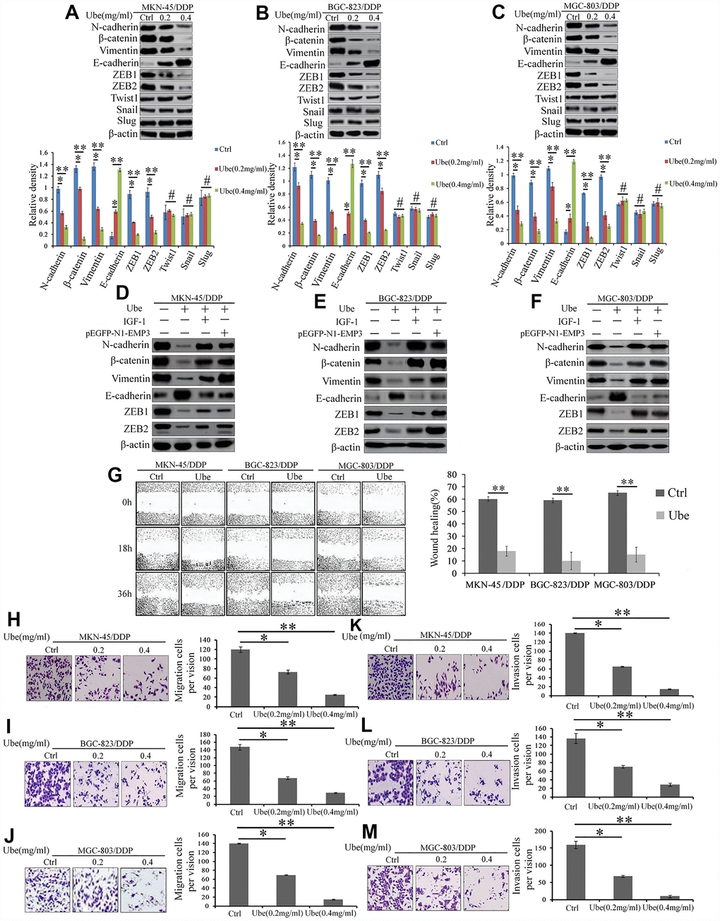
Figure 8.Ubenimex suppresses the EMT, migration, and invasion of CDDP- resistant GC cells by attenuating the activation of the CD13/EMP3/PI3K/AKT/NF-κB pathway. (A–C) Western blot assay was employed to explore the expression of EMT markers in MKN-45/DDP (A), BGC-823/DDP (B) and MGC-803/DDP cells (C), which were stimulated with Ubenimex (0.2 or 0.4 mg/mL) for 24 h. Data are expressed as the representatives (upper panels), and relative expression with means ±SD (bottom panels). **P<0.01,*P<0.05 and #P>0.05. (D–F) Indicated cells were pre-treated with pEGFP-N1-EMP3 plasmid for 24 h or IGF-1 (10ng/mL)or 8h, followed by treatment with Ubenimex (0.4mg/mL) for another 24 h, indicated EMT markers in MKN-45/DDP cells (D), BGC-823/DDP (E) and MGC-803/DDP (F) cells were detected by Western blot assay. (G) Wound healing assays were carried out to determine the migration abilities of CDDP-resistant GC cells which were treated with Ubenimex. Cell morphology of gap with different widths at 0, 18, and 36 h were obtained (left panels), and data are summarized as the means ± SD of “healing ratio”(right panel) **P<0.01. (H–M) Transwell assays was performed to identify the changes of the migration (H–J) and invasive (K–M) abilities in CDDP-resistant GC cells which were treated with Ubenimex (0.2 or 0.4 mg/mL) for 24h. Results were shown as the representatives (left panels) and the number of migrated and invasive cells with the means±SD (right panels) from three experiments. *P<0.05 and **P<0.01.