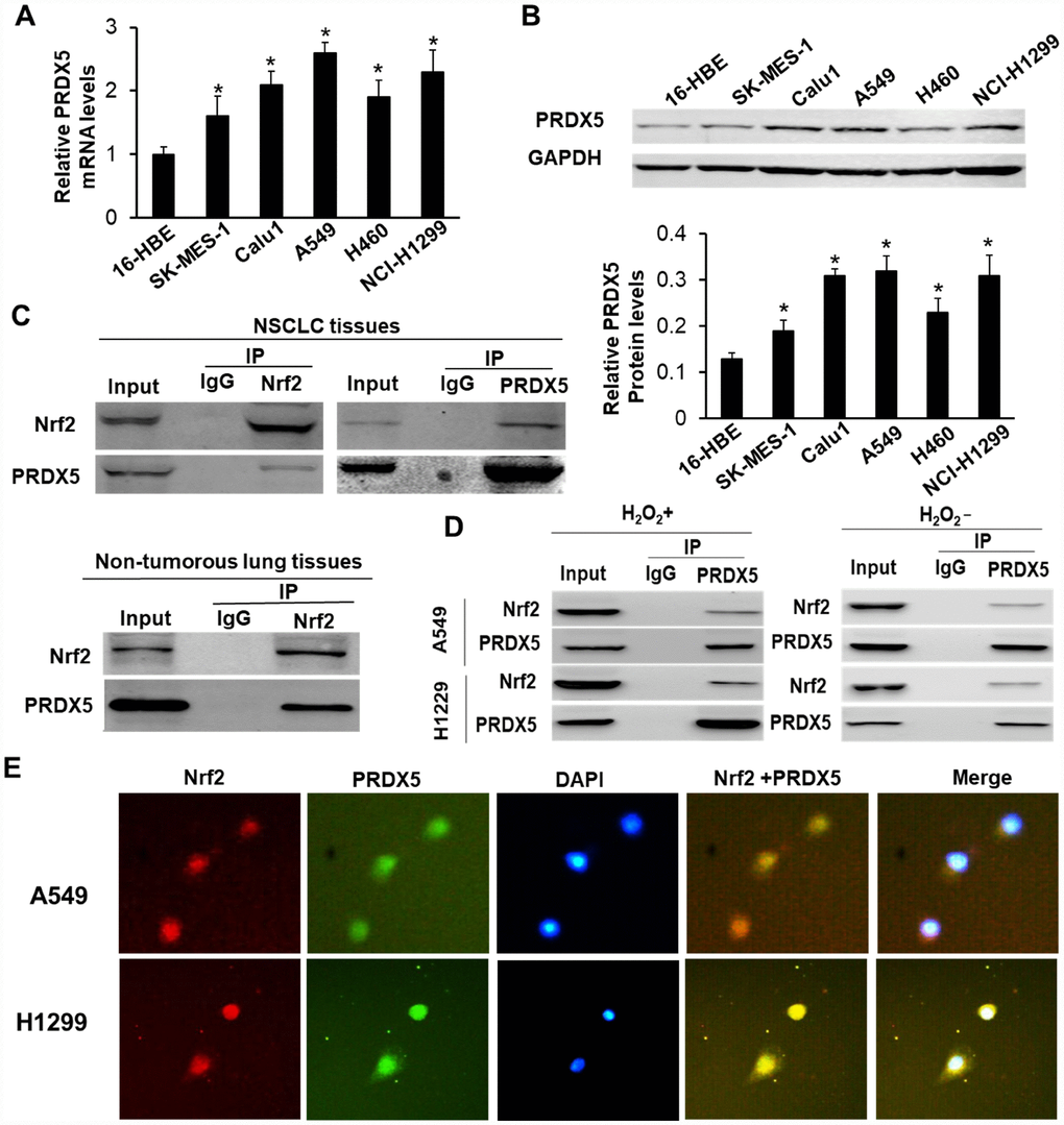
Figure 1.PRDX5 interacted with Nrf2 in NSCLC tissues and related cell lines. (A) qRT-PCR analysis of PRDX5 mRNA level in NSCLC cell lines and 16-HBE cells. The data are reported as the mean ± SD. *P < 0.05, compared with the level in 16-HBE cells. (B) PRDX5 proteins in the different NSCLC cell lines and the normal bronchial epithelial cell 16-HBE analyzed by Western blot analysis. The data shown represent the mean ± SD (*P < 0.05, compared with the level in 16-HBE cells). (C) Reciprocal immunoprecipitation of Nrf2 and PRDX5 in human NSCLC tissue (figure above) and PRDX5 was immunoprecipitated using an anti-Nrf2 antibody in the adjacent normal tissue (figure below). Lysates of the tissues were immunoprecipitated with anti-Nrf2, anti-PRDX5 antibodies or control IgG. The immunoprecipitates were subjected to Western blot analysis with anti-PRDX5 and anti-Nrf2 antibodies. (D) Interaction between Nrf2 and PRDX5 in A549 and NCI-H1299 cells under H2O2 treatment or nontreatment. The lysates obtained from the cells treated with 100 μM H2O2 for 30 min or not were immunoprecipitated using anti-Nrf2, anti-PRDX5 antibodies or control IgG. (E) Immunofluorescence analysis of Nrf2 and PRDX5 in A549 and NCI-H1299 cells. A549 and H1299 cells were pre-incubated with 100 μM H2O2 for 30 min, and then immunostained with a combination of anti-Nrf2 and anti-PRDX5 antibodies. The fluorescent images were digitally merged. Yellow coloration in overlay panels indicates colocalization of Nrf2 and PRDX5. Nuclei were counterstained with DAPI. Scale bar, 50 μm.