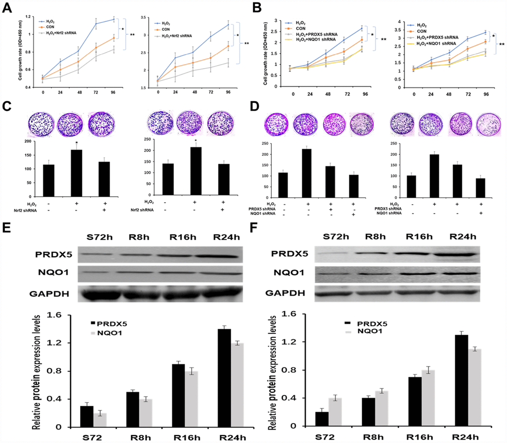
Figure 3.Nrf2 enhanced the growth of NSCLC cells by PRDX5 and NQO1. (A) Growth curves of A549 and H1299 cells treatment with 100 μM H2O2 or knockdown of Nrf2 using CCK-8 assay. All the data are mean ± SD and representative of three independent experiments (*P < 0.05). (B) Effect of PRDX5 or NQO1 knockdown on proliferation of A549 and H1299 cells in the presence of 100 μM H2O2 analyzed by CCK-8 assay. The data are reported as the mean ± SD of three independent experiments (*P < 0.05). (C) Nrf2 had an effect on the colony formation ability of NSCLC cells. Equal numbers of A549 and H1299 cells after treatment as above were seeded onto 6-well plates. The cells were fixed and stained with crystal violet after 14 days. The cell colonies (>0.5 mm in diameter) were counted after staining (mean ± SD, *P < 0.05). (D) Colony-forming capability was measured by colony formation assay in A549 and H1299 cells after transfected with PRDX5 shRNA or NQO1 shRNA and stimulated with 100 μM H2O2. The bar chart showed the number of colonies (>0.5 mm in diameter) in A549 and H1299 cells (mean ± SD, *P < 0.05). (E and F) A549 and H1299 cells were serum starved for 72 h, and then refed with serum for 0, 8, 16 and 24 h. The cell lysates of the corresponding time point were prepared and analyzed by Western blot using antibodies against PRDX5 and NQO1. GAPDH was used as a loading control. The bar charts demonstrated the ratio of PRDX5 or NQO1 to GAPDH by densitometry in A549 and H1299 cells. Mean ± SD of three independent experiments.