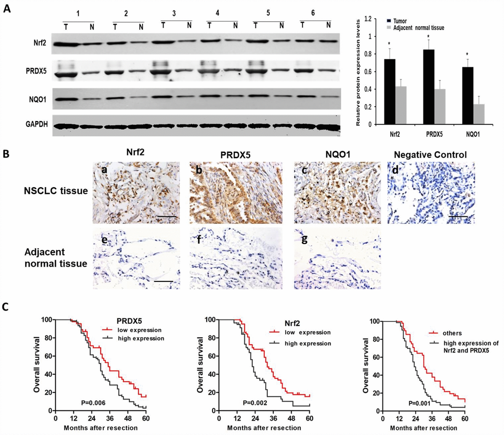
Figure 7.The protein expressions of Nrf2, PRDX5 and NQO1 in NSCLC specimens. (A) The Nrf2, PRDX5 and NQO1 expressions in 26 NSCLC and the adjacent normal tissues by western blot analysis. The representative western blot results in 6 cases are shown. GAPDH was used as a control for protein load and integrity. The bar chart demonstrated the ratio of Nrf2, PRDX5 and NQO1 expression to GAPDH between tumor and non-tumor tissues for the above by densitometry. *,#P < 0.05, significant upregulated expression of Nrf2, PRDX5 and NQO1 in cancerous tissues, compared with adjacent normal tissues. The data are reported as the mean ± SD (*, #P < 0.05, compared with the adjacent tumor tissues). (B) Immunohistochemical analysis of Nrf2, PRDX5 and NQO1 in NSCLC tissues (a–d) and the adjacent normal tissues (e–g). Scale bar, 100 μm. (C) Cumulative survival curves according to Nrf2 and PRDX5 expression in 121 patients with NSCLC. Left, overall survival curves of low PRDX5 expression group vs high PRDX5 expression group (the choosing relative level of 2.323 as the optimal cut-off point of PRDX5). Middle, overall survival curves of low Nrf2 expression group vs high Nrf2 expression group (the choosing relative level of 2.825 as the optimal cut-off point of Nrf2). Right, overall survival curves of high Nrf2/PRDX5 expression group vs the other group.