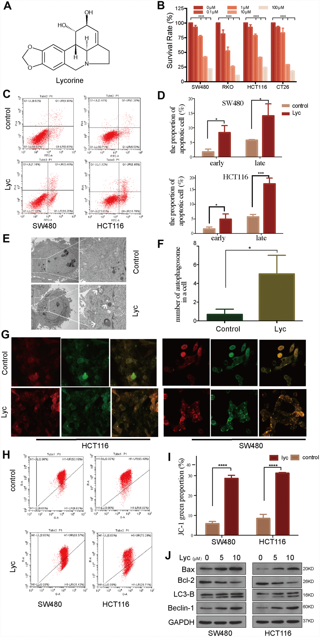
Figure 1.Lycorine induces autophagy-associated apoptosis in colorectal cancer (CRC) cell lines. (A) Chemical structure of lycorine. (B) Four CRC cell lines were treated with the indicated concentrations of lycorine for 24 h. Cell viability was assessed using the Cell Counting Kit-8 assay. (C–D) Cells were treated with lycorine for 24 h and analyzed using annexin V/PI flow cytometry. The right lower quadrant represents early apoptosis. (E–F) The morphological changes in lycorine-treated CRC cells were detected using transmission electron microscopy. Magnification: ×1700 (left), ×5000 (right). (G) HCT116 and SW480 cells were transfected with a tandem fluorescent mRFP-GFP-tagged LC3 virus and then treated with lycorine for 24 h, followed by analysis using confocal fluorescence microscopy (×1000). (H–I) Cells treated with lycorine were harvested, and their mitochondrial membrane potentials were analyzed using a JC-1 kit via flow cytometry. (J) CRC cells were treated with various concentrations of lycorine for 24 h. The apoptosis-related proteins Bax and Bcl-2 and autophagy-related proteins LC3-B and Beclin-1 were analyzed using western blotting. GAPDH was used as a loading control. Data are and presented as the mean ± SD of three independent experiments (*p < 0.05, **p < 0.01, ***p < 0.001, ****p < 0.0001).