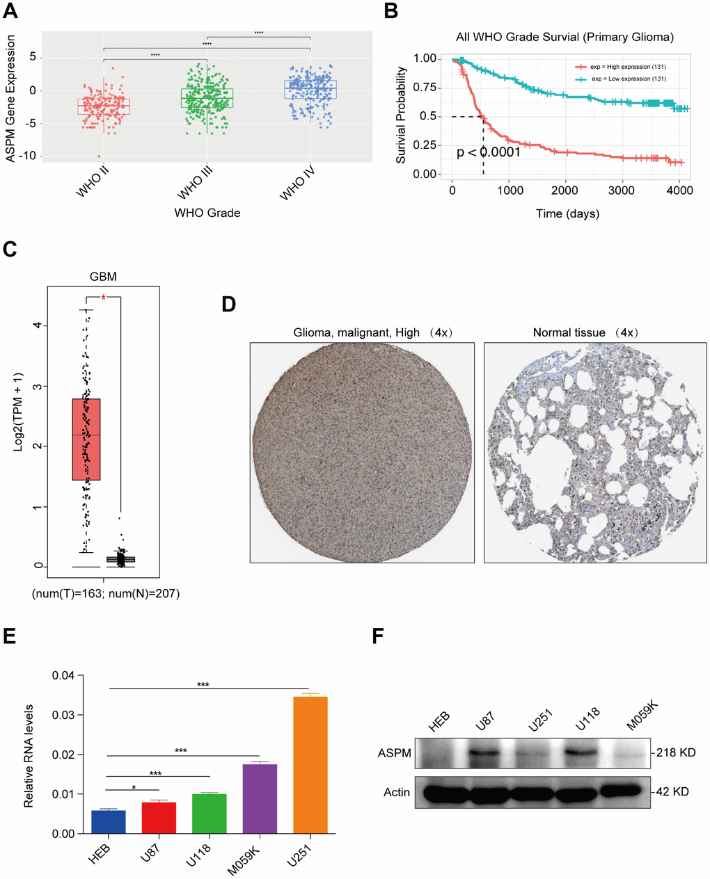
Figure 5.ASPM is highly expressed in human GBM tissues and cell lines. (A) Expression profiles of ASPM in GBM form grade II to grade IV. Red points indicate the expression level of ASPM in grade II glioma; green points indicate the expression level of ASPM in grade III glioma, and blue points indicates the expression level of ASPM in grade IV glioma. (B) Patients with high expression of ASPM in glioma had a significantly poor prognosis (n=262, p < 0.0001). (C) Box plot of ASPM expression in GBM from GEPIA. The box plot is based on 163 GBM cancer samples and 207 normal samples. The expression level of RNA was upregulated in GBM tissues compared with normal tissues. *p < 0.05 indicates a significant difference. (D) The Human Protein Atlas project shows representative immunohistochemical images of ASPM in GBM compared with normal tissues. (E) and (F) The RNA and protein levels of ASPM were detected in four GBM and one normal glial cell line by RT-qPCR and western blot. *p < 0.05, *** p < 0.001.