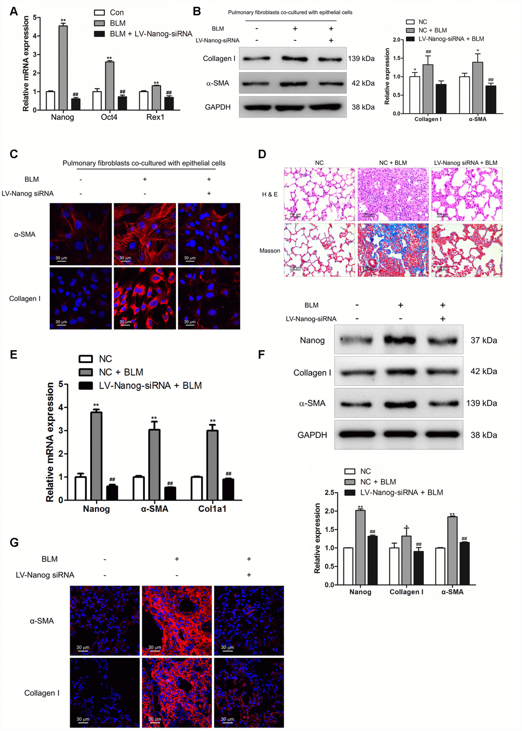
Figure 6.Nanog silencing could suppress pulmonary fibroblast activation and impair the development of pulmonary fibrosis. (A–C) Pulmonary fibroblasts were transfected with LV-Nanog-siRNA and co-cultured with MLE-12 cells as in Figure 3D. (A) The mRNA levels of Nanog, Oct4 and Rex1 were measured by Q-PCR, ** P < 0.01 vs. Con and ## P < 0.01 vs. bleomycin (BLM). (B) The protein levels of collagen I and α-SMA were determined by Western blot. The expression levels were quantified with ImageJ (n = 3). GAPDH was used as a loading control, *P < 0.05 and **P < 0.01. (C) The expression of collagen I and α-SMA were further examined by immunofluorescence staining. (D–G) Mice were intratracheally injected with 5 × 10⁸ TU/ml LV-Nanog-siRNA or negative control (NC) 7 days after administration of BLM. Mice were sacrificed on day 21 after BLM instillation. (D) Pulmonary fibrosis was determined by haematoxylin and eosin (H&E) staining and collagen I was revealed by Sirius Red/Fast Green staining. (E) The mRNA levels of Nanog, α-SMA and collagen I were determined by Q-PCR, ** P < 0.01 vs. NC and ## P < 0.01 vs. NC + BLM. (F) The protein levels of Nanog, collagen I and α-SMA were measured by Western blot. The expression levels were quantified with ImageJ (n = 3). GAPDH was used as a loading control, *P < 0.05 and **P < 0.01. (G) The expression of α-SMA and collagen I were further confirmed by immunofluorescence staining.