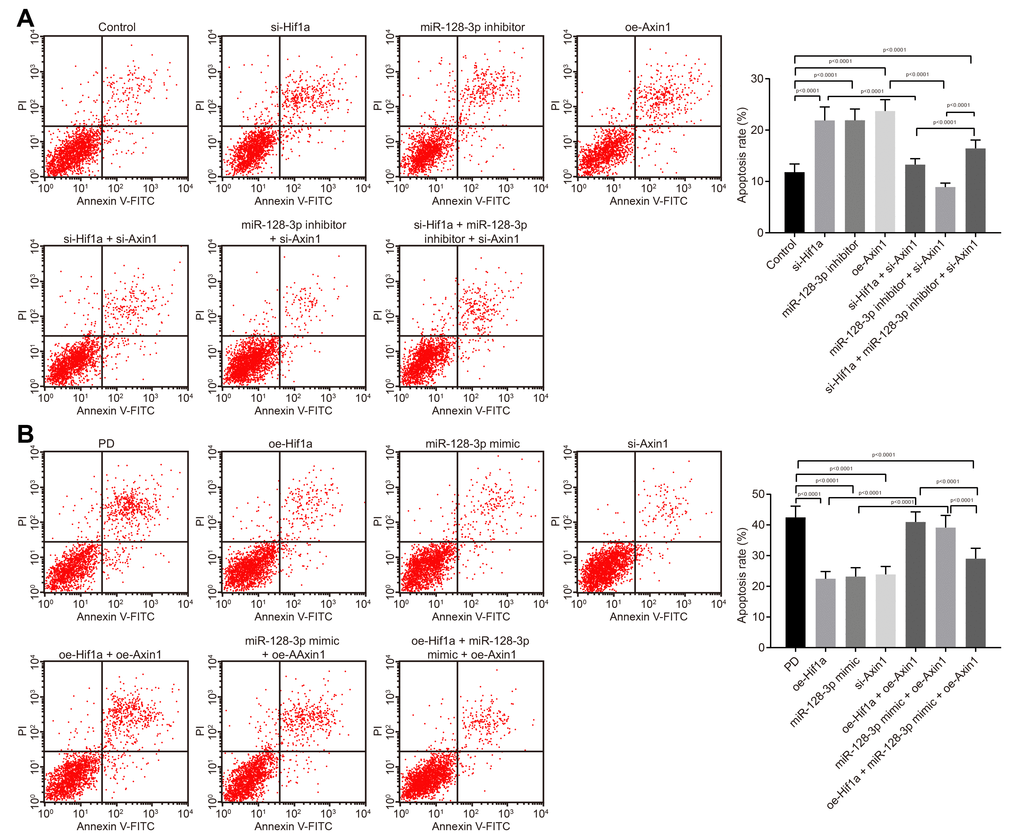
Figure 4.The apoptosis of hippocampal neurons cultured from normal mice and an MPTP-lesioned mouse model of PD mediated by the HIF-1α/miR-128-3p/AXIN1 axis. (A) The apoptosis of hippocampal neurons of normal mice after treatment with si-Hif1a, miR-128-3p inhibitor, or si-Axin1 as detected by flow cytometry. * p < 0.05 vs. the control group (primary hippocampal neurons of normal mice); # p < 0.05 vs. the si-Hif1a group (hippocampal neurons of normal mice treated with si-Hif1a); & p < 0.05 vs. the miR-128-3p inhibitor group (hippocampal neurons of normal mice treated with miR-128-3p inhibitor); @ p < 0.05 vs. the si-Hif1a + si-Axin1 group (hippocampal neurons of normal mice treated with si-Hif1a + si-Axin1); $ p < 0.05 vs. the miR-128-3p inhibitor + si-Axin1 group (hippocampal neurons of normal mice treated with miR-128-3p inhibitor + si-Axin1). (B) The apoptosis of hippocampal neurons in the MPTP-lesioned mouse model of PD after treatment with oe-Hif1a, miR-128-3p mimic, or oe-Axin1 detected by flow cytometry. * p < 0.05 vs. the PD group (primary hippocampal neurons of the MPTP-lesioned mouse model of PD); # p < 0.05 vs. the oe-Hif1a group (hippocampal neurons of the MPTP-lesioned mouse model of PD treated with oe-Hif1a); & p < 0.05 vs. the miR-128-3p mimic group (hippocampal neurons of MPTP-lesioned mouse model of PD treated with miR-128-3p mimic); @ p < 0.05 vs. the oe-Hif1a + oe-Axin1 group (hippocampal neurons of MPTP-lesioned mouse model of PD treated with oe-Hif1a + oe-Axin1); $ p < 0.05 vs. the miR-128-3p mimic + oe-Axin1 group (hippocampal neurons of MPTP-lesioned mouse model of PD treated with miR-128-3p mimic + oe-Axin1). The experiment was repeated three times independently.