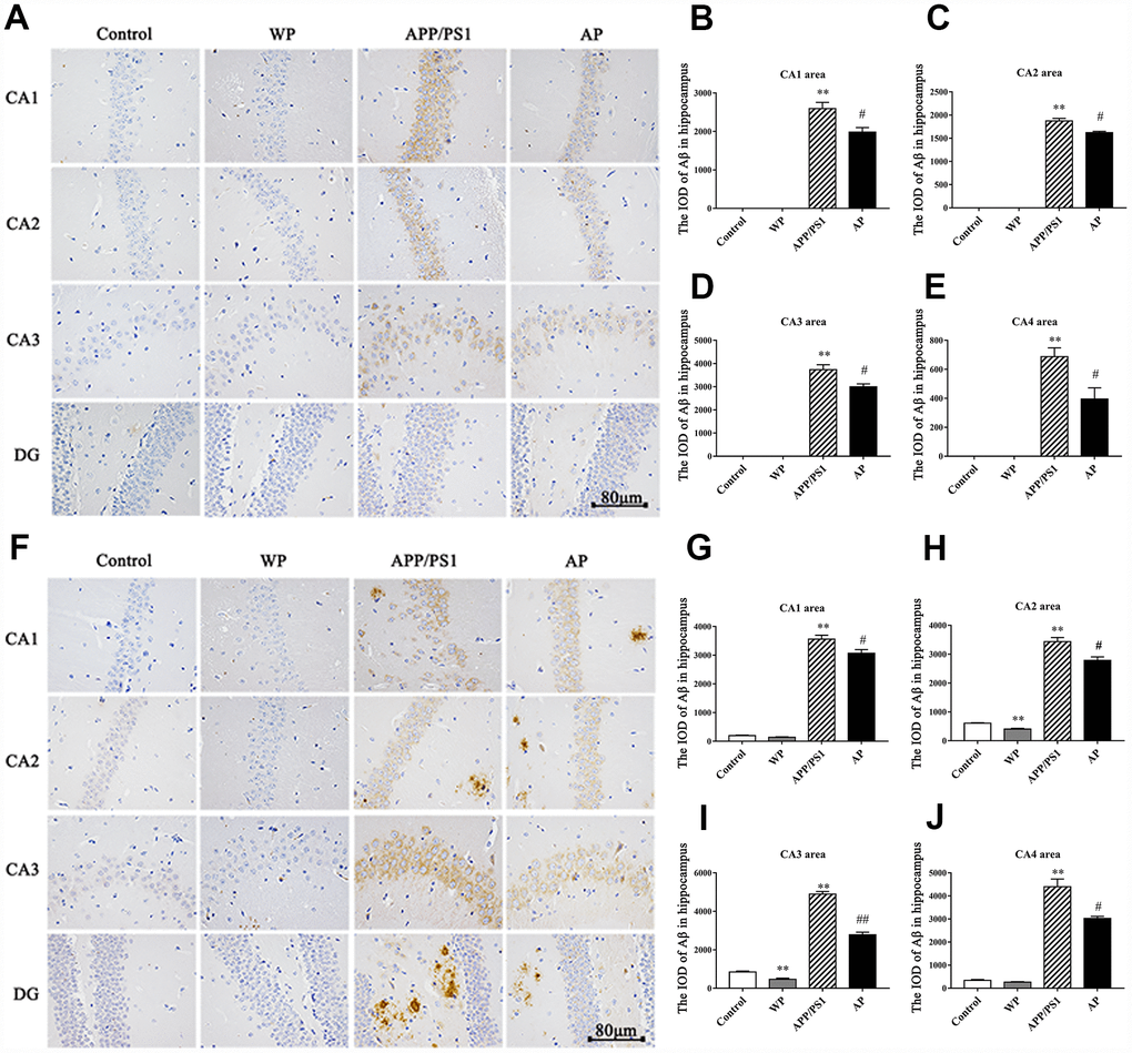
Figure 3.Activation of α7 nAChR reduces the deposition of Aβ in the hippocampus of the APP/PS1_DT mice. The deposition of Aβ in the hippocampus of APP/PS1_DT mice (A) 6 months old and (F) 10 months old. The IOD of Aβ in the hippocampus area of 6 months old mice in the (B) CA1 area, (C) CA2 area, (D) CA3 area, (E) CA4 area, and 10 months old mice in the (G) CA1 area, (H) CA2 area, (I) CA3 area and (J) CA4 area. Control, WT C57 mice injected with saline; WP, WT mice injected with PNU; APP/PS1, APP/PS1_DT mice injected with saline; AP, APP/PS1_DT mice injected with PNU. CA1, CA2, CA3 and DG indicate the CA1 area, CA2 area, CA3 area and DG area of the hippocampus, respectively. Compared with the control group, Aβ deposition in the hippocampus of APP/PS1_DT mice was increased significantly, and this trend was partially reversed by PNU treatment (AP group). The results demonstrated that α7 nAChR partially reduced the deposition of Aβ in the hippocampus of the APP/PS1_DT mice. Data are presented as the mean ± standard deviation. *P<0.05, **P<0.01 vs. control group; #P<0.05, ##P<0.01 vs. APP/PS1 group.