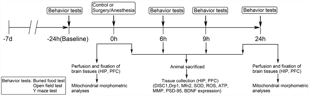
Figure 1.Diagram of the experimental design. The mice underwent behavioral tests at 24 hours before (baseline) and at 6, 9, and 24 hours after the Surgery/Anesthesia. Within each group, separate cohorts were subjected to assessments at each time point (n = 9 per cohort). Mice were sacrificed immediately after the Surgery/Anesthesia and at 6, 9, and 24 hours postoperatively. The hippocampal and prefrontal cortex tissues were harvested for analysis of DISC1, Drp1, Mfn2, SOD, ROS, ATP, MMP, BDNF, and PSD-95 levels (n = 6 per cohort). Mice were anesthetized and transcardially perfused with ice-cold phosphate-buffered saline (PBS) followed by paraformaldehyde and glutaraldehyde; then, hippocampal and prefrontal cortex tissues were collected and stored in the same fixative for electron microscopy analysis immediately after the Surgery/Anesthesia and at 24 hours postoperatively (n = 3 per cohort). DISC1, disrupted in schizophrenia 1. Drp1, dynamin-related protein 1. Mfn2, mitofusin 2. SOD, superoxide dismutase. ROS, reactive oxygen species. ATP, adenosine triphosphate. MMP, mitochondrial membrane potential. PSD-95, postsynaptic density protein 95. BDNF, brain-derived neurotrophic factor.