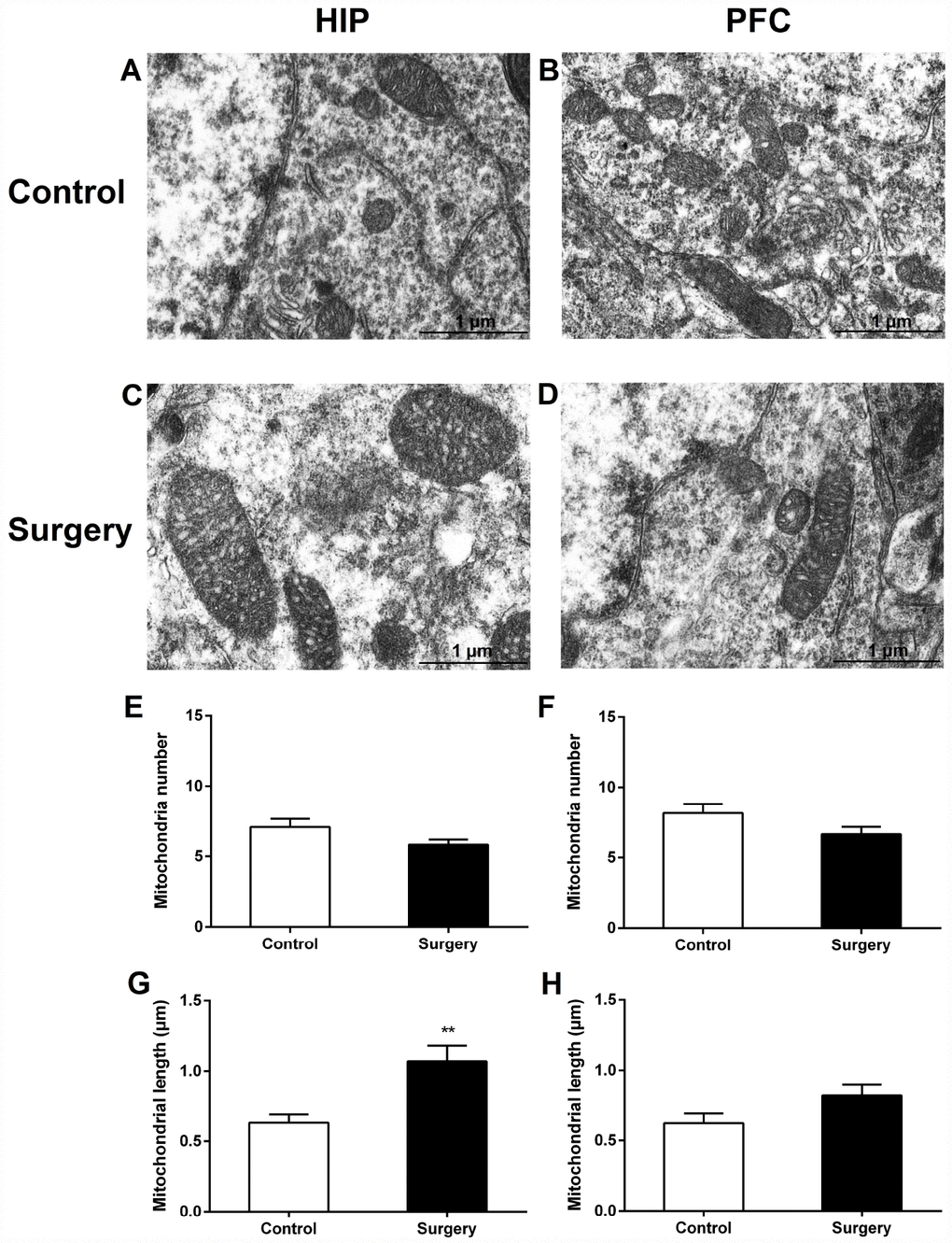
Figure 6.Surgery/Anesthesia caused acute ultrastructural changes in the mitochondria of hippocampal but not prefrontal cortex neurons in aged mice immediately after Surgery/Anesthesia. Mitochondria in the cytoplasm of hippocampal (A) and prefrontal cortex (B) neurons from the control mice resemble long tubules with intact outer and inner membranes and numerous cristae tightly packed in healthy looking matrix. Compared with those in the control group, mitochondria in the cytoplasm of hippocampal neurons (C) from mice in the Surgery/Anesthesia group became swollen, while the ultrastructure of mitochondria in the prefrontal cortex neurons (D) was normal immediately after Surgery/Anesthesia. The number and length of mitochondria were measured in the hippocampus (E, G) and prefrontal cortex (F, H) in 6 different fields of view per animal. (G) Surgery/Anesthesia increased mitochondrial length in the hippocampus compared to the control condition at 0 hour postoperatively. Scale bar: 1 μm. The data are plotted as the mean ± standard error of the mean for each group (n = 3). **p < 0.01, compared to control.