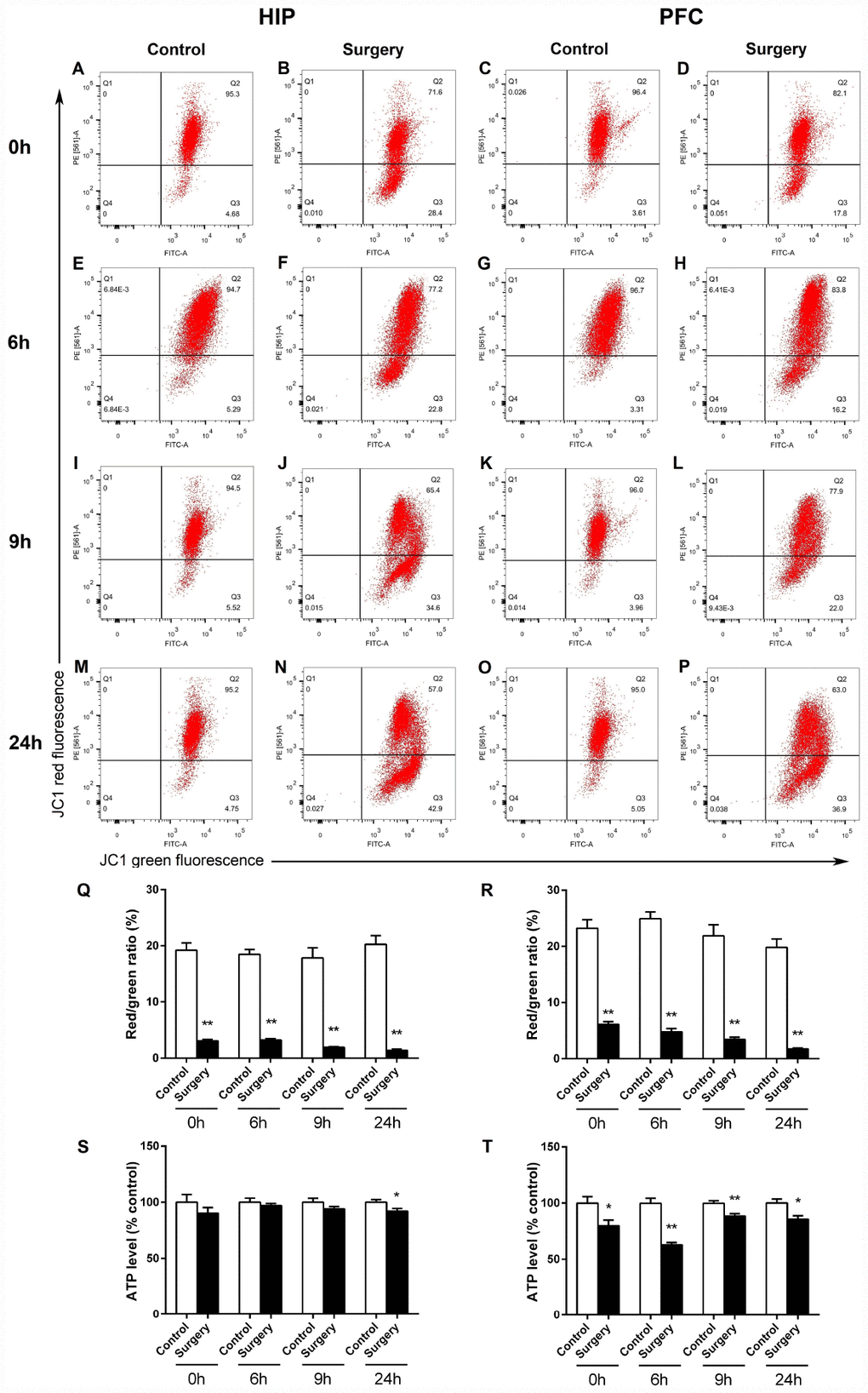
Figure 9.Surgery/Anesthesia altered the levels of MMP and ATP in the hippocampus and prefrontal cortex of aged mice at 0, 6, 9, and 24 hours postoperatively. Changes in MMP were measured using flow cytometry and JC-1. Representative graphs of flow cytometric analysis of the altered MMP level in the hippocampus (A, B, E, F, I, J, M, N) and prefrontal cortex (C, D, G, H, K, L, O, P) of mice after incubation with JC-1. Statistical bar graphs show the changes of MMP detected using flow cytometry. The changes of MMP in the hippocampal (Q) and prefrontal cortex tissues (R) were defined as the ratio of red/green fluorescence intensity. Surgery/Anesthesia reduced the MMP level in the hippocampus and prefrontal cortex as compared to the control condition in mice immediately after Surgery/ Anesthesia. (S) Surgery/Anesthesia decreased the ATP level in the hippocampus at 24 hours but not at 0, 6, or 9 hours postoperatively. (T) In the prefrontal cortex, Surgery/Anesthesia significantly decreased the level of ATP as compared to control condition in mice at all the postoperative timepoints. The data are plotted as the mean ± standard error of the mean for each group (n = 6). *p < 0.05 and **p < 0.01, compared to control.