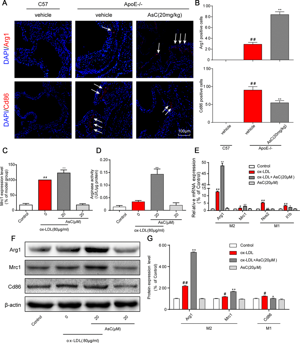
Figure 3.AsC polarized macrophages to an M2-like phenotype. All mice were fed a HFD in the presence or absence of AsC (20 mg·kg-1·day-1, i.g.) for 4 weeks. In the in vitro assay, RAW264.7 cells were pretreated with AsC (20 μM) for 12 h, and then exposed to ox-LDL for another 24 h. (A) Dual immunofluorescence staining for Arg1 (red) or Cd86 (red) and DAPI (blue) in lesions in the aortic root. (B) Quantification of the relative fluorescence intensity. (C) The Mrc1 expression level in ox-LDL-treated macrophages, as determined by flow cytometry. (D) Arginase activity was measured as described in the Methods section. (E) mRNA levels of Arg1, Mrc1, Nos2 and Il1b in macrophages, as quantified by real-time PCR. (F) Representative photographs of Mrc1, Cd86 and Arg1 expression in ox-LDL-treated macrophages, as evaluated by western blot analysis. (G) Statistical results of Mrc1, Cd86 and Arg1 expression levels compared with those in the control group. The data are presented as the means ± SDs (n = 5). #P < 0.05, ##P < 0.01 vs. the control group, **P < 0.01 vs. the model group.