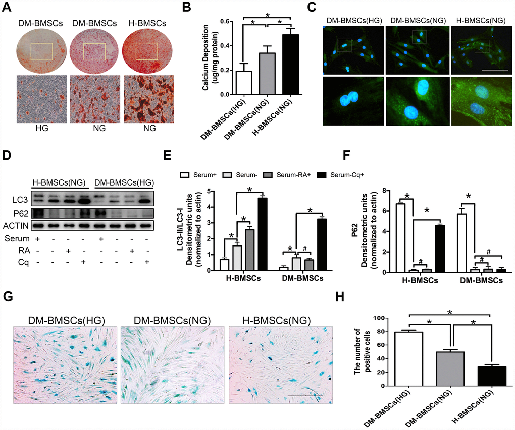
Figure 1.DM-BMSCs show a decreased osteogenic differentiation and autophagy level, and an increased senescent phenotype. DM-BMSCs and H-BMSCS were incubated in the corresponding condition and treated by osteogenic inducement for 14 days and stained with Alizarin Red S (A). Quantitative analysis of the amount of calcium-bound stain was determined by comparison with calcium standards (B). Fluorescence detection of autophagosomes in DM-BMSCs and H-BMSCs transfected with the GFP-LC3 plasmid and cultured in serum deprivation conditions for 6 h (C). The expression of LC3 and P62 were detected by western blot after serum deprivation for 6 h (D). Protein bands were quantified and analyzed by densitometric analysis. Rapamycin (RA) as positive control and chloroquine (Cq) as negative control (E, F). Cell senescence was detected by SA-β-Gal staining after incubation in the corresponding condition (G). The number of positive cells was calculated (H). NG, normoglycemic condition, HG, hyperglycemic condition. Data are presented as the mean ± standard deviation, n=3. *p<0.05, #p>0.05. Scale bar = 100 μm.