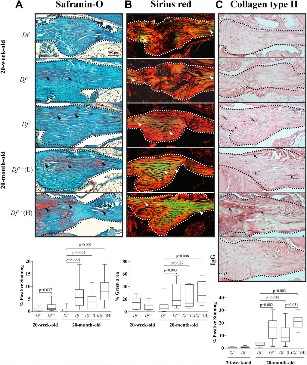
Figure 2.Histology and type II collagen deposition in the anterior cruciate ligament (ACL). Photomicrographs of representative images and box plots of 20-week-old adipsin-deficient (Df-/-) and wild-type (Df+/+) and 20-month-old Df-/-, Df+/+, Df+/+(L) and (H) mice of (A) Safranin-O staining, black arrowheads indicate proteoglycans deposition; (B) Sirius red staining enabling visualization of the collagen fibers. The green fibers corresponding to altered collagen were quantified over the total area. White arrowheads indicate thin collagen fibers. (C) Immunohistochemistry of type II collagen deposition and a negative control (IgG) performed by substitution with a non-specific rabbit IgG. Black arrows indicate positive staining. In (A–C) dotted lines delineate the core portion of the ACL. Bar in (A) = 100 μm. Original magnification X100. Values are the median and interquartile range of Df-/- (n=11), Df+/+ (n=13) for the 20-week-old mice and of Df-/- (n=13), Df+/+ (n=13), Df+/+ (L) (n=7) and (H) (n=6) for the 20-month-old mice. p values were determined by the Mann-Whitney test. Only significant differences are shown except for those comparing 20-week-old and 20-month-old Df-/- (C, p= 0.0001) and Df+/+ (A, p= 0.006; B, p=0.004; C, p< 0.0001) mice.