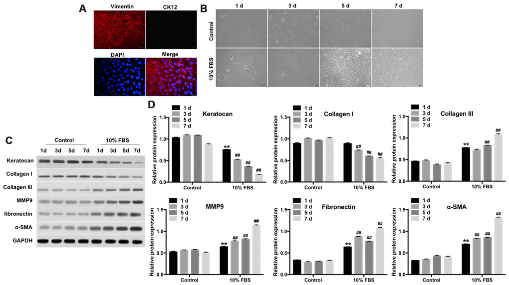
Figure 1.FBS induces differentiation of rabbit corneal keratocytes into myofibroblasts. (A) Representative immunofluorescence images show staining of rabbit corneal stromal cells with fluorescent-tagged antibodies against Vimentin (red) and CK12 (green). Vimentin-positive cells are the keratocytes; CK12-positive cells are the corneal epithelial cells; Diamidinophenylindole (DAPI; blue) stains the nucleus; magnification: 200x. (B) Representative phase-contrast images show phenotypic features of rabbit corneal keratocytes cultured in 10% FBS or serum-free DMEM/F12 medium for 1, 3, 5 and 7 days. The cell cultures were observed under a light microscope and images were captured at 10X magnification. (C) Representative western blot images show levels of keratocan, collagen I, collagen III, MMP9, fibronectin and α-SMA proteins in rabbit keratocyte cells grown in DMEM/F12 medium containing 10% FBS or serum-free medium for 7 days. GAPDH was used as an internal control. (D) Histogram plot shows keratocan, collagen I, collagen III, MMP9, fibronectin and α-SMA protein levels relative to GAPDH. ** denotes P < 0.01 compared with the control (1d) group; ## denotes P < 0.01 compared with the 10% FBS (1d) group.