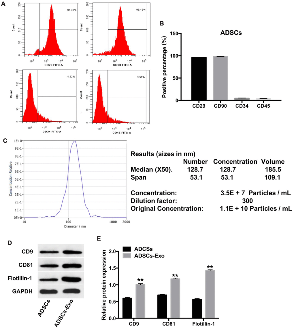
Figure 2.Characterization of ADSCs and ADSCs-Exo. (A, B) Flow cytometry analysis of ADSCs isolated from rabbit adipose tissues is shown using fluorescent-tagged antibodies against cell surface proteins, namely, CD29, CD90, CD34, and CD45. (C) The mean diameter of ADSCs exosomes was analyzed using a nanoparticle tracking system (NTA). NTA analysis of the exosomes isolated from ADSCs (ADSCs-Exo) shows a mean concentration of 1.1 x 10¹⁰ particles per mL. (D, E) Western blot analysis shows levels of CD9, CD81 and flotillin-1 proteins in the ADSCs and the ADSCs-Exo. GAPDH was used as an internal control. The levels of CD9, CD81 and flotillin-1 are expressed relative to GAPDH. ** denotes P < 0.01 compared with the ADSC group.