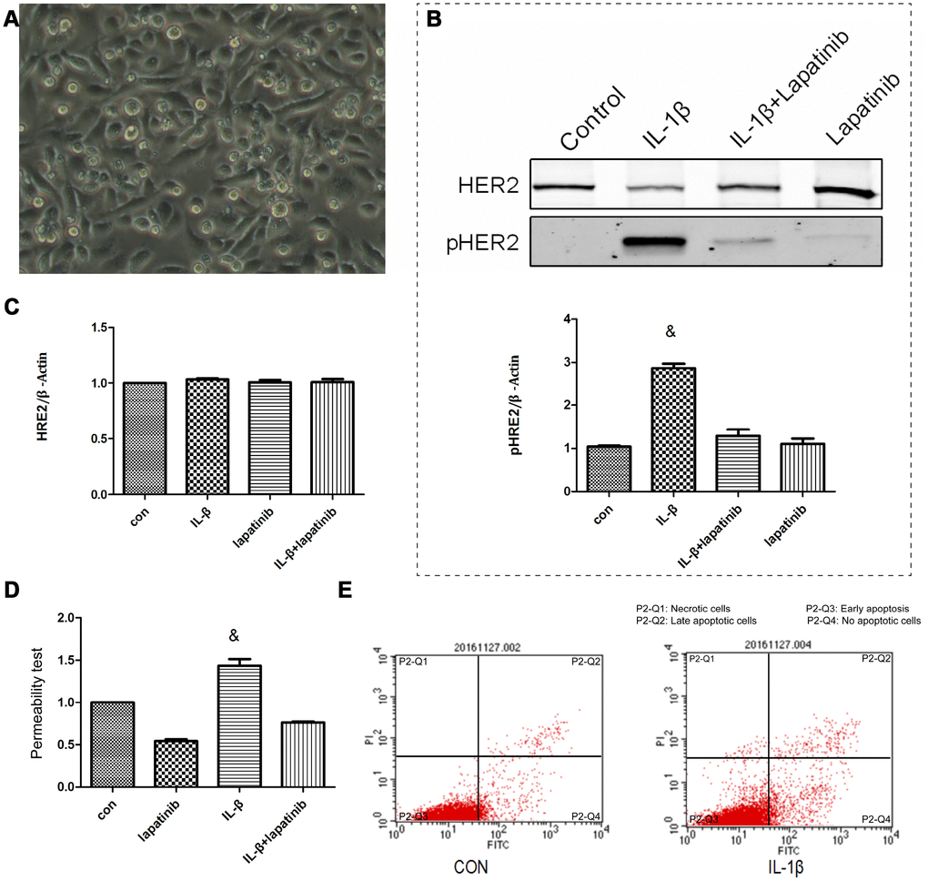
Figure 3.The lung epithelial cells were divided into 4 groups. Control group, IL-β group, lapatinib group and IL-β+lapatinib group. (A) Human lung epithelial cells were cultured in vitro, and the cells were adherently grown in culture flasks. Generally, the growth was good, the nucleus was located in the center of the cells, and the cytoplasm was extended outward. Once polygons grow irregularly. (B) The pHER2 were detected in lung epithelial cells using western blot method. Compared with the control group, the expression of pHER2 was significantly up-regulated in IL-1β group. &, P < 0.01. (C) The expression of HER2 was not significantly different in the four groups. (D) Compared with the control group, the permeability of epithelial cells increased after 6 hours of IL-1β treatment. There was no significant difference between the other groups. &, P < 0.01. (E) Under the action of IL-1β, there was no increase in apoptosis.