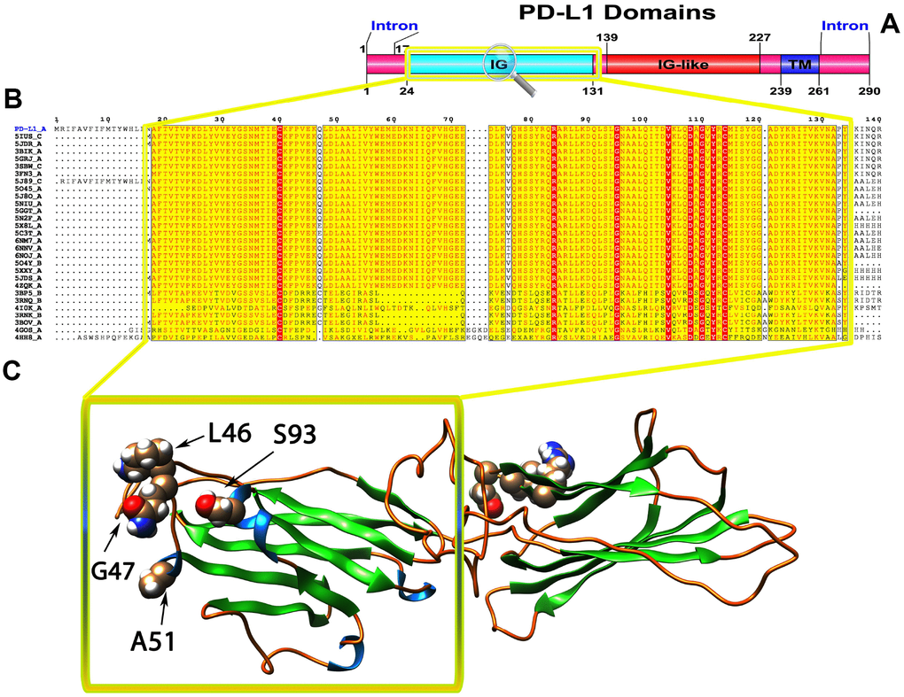
Figure 2.(A) Molecular structure and Conserved domain analysis of PD-L1 protein. (B) Showing the MSA of the 20 most homologous proteins to PD-L1 (obtained with a BLAST+ search against the PDBAA database). Known secondary structure elements are displayed for all aligned sequences. Alternate residues are highlighted by gray. Identical and similar residues are boxed in red and yellow, respectively. (C) Location of positively selected amino acid sites identified PD-L1 conserved Ig domain. The crystal structure of human PD-L1 was used as a reference sequence and positively selected sites were drawn onto the crystal structure using Phyre tool (http://www.sbg.bio.ic.ac.uk/ phyre2/html). Four residues identified under selection fall in the immunoglobulin-like domain containing the ligand-binding site. The sites which fall in the region identified as the ligand-binding site and another cluster in a region immediately following the signal sequence.