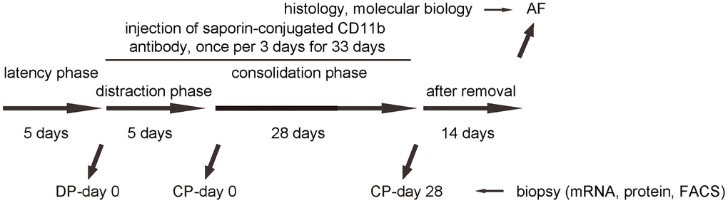
Figure 3.Analysis of biopsy tissue during DO with macrophage depletion. Illustration of the model: We performed DO of the lower limb on the C57/Bl6 mice. After surgery, there was a 5 days’ latency for the mice the recover. Afterwards, there was a 5 days’ distraction phase constituted by a rate of 0.5 mm’ distraction per 24 hours. After distraction phase, a 28 days’ consolidation phase was applied, followed by removal of external fixers and a 14 days’ delay for final analysis. In order to analyze macrophages during DO-induced bone regeneration, we took biopsy of the tissue in the surgical/regeneration area at day 0 of distraction phase (DP-day 0), day 0 of consolidation phase (CP-day 0) and day 28 or consolidation phase (CP-day 28). In order to understand the role and necessity of macrophages in DO-induced bone regeneration, we performed an interference by inducing macrophage depletion during DO. DO-surgery-treated mice received i.v. injection of either saporin-CD11b once every 3 days, or control rat IgG of same frequency (IgG). The injection started at DP-day 0 and ended at CP-day 28.