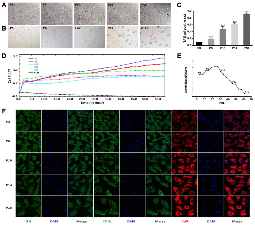
Figure 1.Characterization of senescence in HUVEC cells during continual passaging. (A) Cell morphological characteristics (×100 magnification). (B) Senescence-associated-galactosidase (SA-β-gal) staining (×100 magnification). (C) Percentages of SA-β-gal-positive cells. Data were presented by means ± SE, *** denotes the statistical significance p < 0.001 relative to the P3 group. (D) Real-time cell growth curves. The initial 4 or 5 h was the time for cell adherence. (E) Cell growth rates. (F) Cell immunofluorescence assay (×400 magnification). Positive immunoreactivity is shown for several endothelial markers including anti-F8 (green), CD31 (green) and von Willebrand factor (vWF; red) antibodies. Nuclei were stained with 4’, 6-diamidino-2-phenylindole (DAPI) as a contrast (blue).