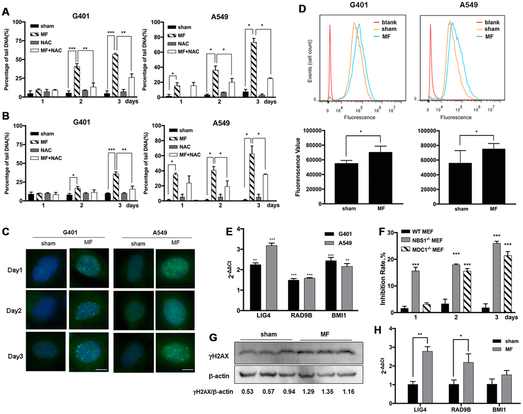
Figure 5.ROS-induced DNA damage and activation of DNA repair pathways following MF exposure. G401 and A549 cells were subjected to MF or sham exposure, 2 h daily for 3 consecutive days, with or without incubation with NAC (1 mM). (A, B) Percentage of tail DNA detected by alkaline (A) and neutral (B) Comet assays. (C) Subcellular localization of γH2AX in G401 and A549 cells. Scale bar= 5 μm. (D) Expression of DNA-PKCs protein in G401 and A549 cells with MF exposure (MF) or sham exposure (sham) on day 2, detected by flow cytometry analysis (n=3). *: P<0.05. (E) mRNA expression of genes in DNA repair system including LIG4, RAD9B and BMI1 in G401 and A549 cells. Asterisk indicates comparison with sham exposure (n=3). (F) WT and MDC1 or NBS1 deficient MEF cells were subjected to the same MF exposure protocol. Inhibition rates were calculated from number of viable cells. Data are expressed as mean ± SE from 3 independent experiments (n=5 in each experiment). Asterisk indicates comparison with WT-MEF. (G, H) G401 nephroblastoma was established in nude mice. Expression of γH2AX protein (G) and selected genes from the DNA repair system (H) are shown (n=3). *: P<0.05; **: P<0.01; ***: P<0.001.