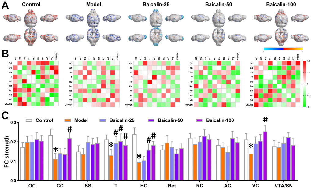
Figure 4.Oral Baicalin supplementation restores brain functional connectivity in repeated cerebral ischemia-reperfusion model mice. (A) Virtual graphics show brain functional connectivity (FC) in control, model, and baicalin-treated model group mice. (B) The mean FC matrices show the strength of functional connectivity between pairs of brain regions in control, model, and baicalin-treated model group mice. The color scale represents the strength of the functional connectivity. (C) The mean functional connectivity strength per brain network for control, model, and baicalin-treated model group mice is shown. Note: * denotes P<0.05 compared with the control mice using unpaired Student`s t-tests; # denotes P<0.05 compared with the model mice using two-way repeated-measures ANOVA with post-hoc Tukey multiple comparisons test. All the values are expressed as means ± S.D. Each group had 15 mice (n=15). The brain regions analyzed include orbitofrontal cortex (OC), cingulate cortex (CC), somatosensory cortex (SS), thalamus (T), hippocampus (H), retrosplenial cortex (Ret), rhinal cortex (RC), auditory cortex (AC), visual cortex (VC), and ventral tegmental area/substantia nigra (VTA/SN).