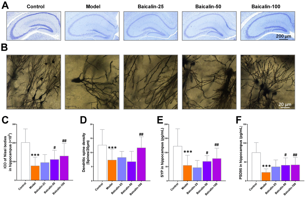
Figure 5.Baicalin improves hippocampal neuronal plasticity in repeated cerebral ischemia-reperfusion model mice. (A, B) Representative confocal microscopic images show (A) Nissl-stained and (B) golgi-stained hippocampal region of the brains of control, model, and Baicalin-treated model group mice. The brain tissue sections were subjected to Nissl and golgi staining, respectively. (C) The total number of Nissl bodies and (D) mean dendritic spine density of the hippocampal neurons based on the analysis of Nissl- and golgi-stained brain tissue sections of control, model, and Baicalin-treated model group mice are shown. (E, F) Representative images show immunohistochemical staining of (E) synaptophysin and (F) PSD95 proteins in the hippocampal region of control, model, and Baicalin-treated model group mice are shown. Note: *** denotes P<0.001 compared to the control mice using unpaired Student`s t-tests; # denotes P<0.05 and ## denotes P<0.01 compared to the model mice using one-way ANOVA and the Dunnett`s post hoc test or a two-way repeated-measures ANOVA and the post-hoc Tukey multiple comparisons test. All the values are expressed as means ± S.D. Each group had 15 mice (n=15).