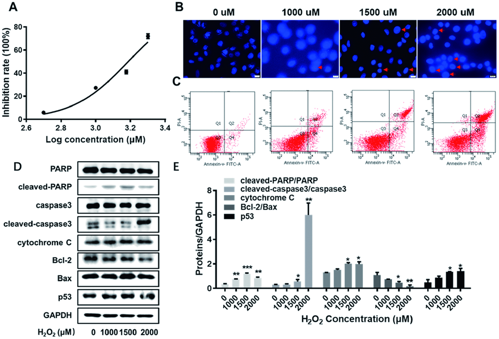
Figure 2.The establishment of the lesion model for HUVECs. (A) The inhibition rate of HUVECs exposed to H2O2 at varied concentrations (0, 500, 1000, 1500 and 2000 uM) were detected by CCK8 assay. (B) HUVECs exposed to H2O2 at varied concentrations (0, 1000, 1500 and 2000 uM) were visualized according to Hoechst staining kit assay. The red arrows indicate nuclear condensation or apoptosis. (C) The apoptosis rate of HUVECs exposed to H2O2 at varied concentrations (0, 1000, 1500 and 2000 uM) were detected by flow cytometry assay. (D) With the enhanced concentrations of H2O2 (0, 1000, 1500 and 2000 uM), the expression levels of cleaved-PARP/PARP, cleaved-caspase3/caspase3, cytochrome C, Bcl-2, Bax and p53 were detected by western blot assay. (E) The corresponding grey-scale maps of (D) were shown. *, P<0.05, **, P<0.01, ***, P<0.001. HUVECs, human umbilical vein endothelial cells; GAPDH, glyceraldehyde 3-phosphate dehydrogenase.